


1. Introduction
With the development of medical technology and science, there are corresponding solutions for many diseases, such as taking medicine, surgery and so on. The brain is one of the most complex and important organs in the human body. In other words, the brain controls most things in the human body, such as muscle movement, thinking, memory, etc. Research on brain diseases is also very important. However, since the brain is relatively fragile, When the brain is infected with a disease, the mortality rate is very high, and even with timely and effective treatment, sequelae will still remain.
As the population ages, various diseases are more likely to occur in the elderly. Stroke is one of these diseases. Stroke is a common disease after the age of 55. After the age of 55, the probability of contracting a stroke doubles every 10 years [1]. And because the immunity of the elderly is reduced, the mortality rate of stroke among the elderly is very high. There are two main types of strokes, each with a different brain injury. An ischemic stroke is when part of the brain does not get enough blood, which dies. That part of the brain might control movement, speech, or thinking. The other type of stroke, a haemorrhagic stroke, is when a blood vessel ruptures in the brain, which puts too much pressure on brain cells, which damages them.
With the development of artificial intelligence (AI) in recent years, AI will also be involved in the treatment of diseases. Due to the intervention of AI, the mortality rate of stroke is now much lower than before, but stroke still causes irreversible damage to the brain, thus affecting normal life. The demand for rehabilitation is still high. The Brain Machine Interface (BMI) plays a big role in rehabilitation. BMI can detect and receive signals from the brain and transmit them to a computer. Scientists or computers can then extract information from these signals [2]. This will include information such as blood pressure and brain activity that can determine whether a stroke has occurred or the possibility of a stroke.
This article mainly analyzes the causes, effects, and AI's prediction and treatment of the two types of strokes. Stroke and AI are both topics that are currently receiving a lot of attention. When AI and BMI are combined, some new topics are provided.
2. Types and characteristics of strokes
With the development of medical technology and science, people have made progress in exploring the brain. Some brain diseases can also be treated. Stroke, a common brain disease among the elderly, has also received attention. There are two main strokes, ischemic and haemorrhagic. These two strokes have different causes, but both damage the brain.
The brain is the most important, fragile, and complex organ in the human body. It mainly controls various functions of the human body, such as movement, speech, behavior, thinking, etc.
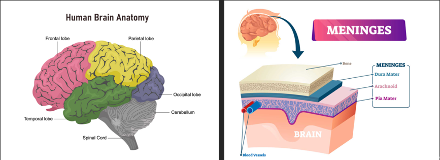
Figure 1: Parts of the brain [3]
Figure 1 shows the parts of the brain. The human head has many layers of structure. The outermost layer is the skull, which is responsible for protecting the brain. The second is the meninges. The meninges have three layers of structure, which are mainly responsible for distributing blood vessels. The blood vessels then provide oxygen to the cells in the brain.
The human brain can be divided into four main parts, each with a different function. The largest part of the brain is the frontal lobe which is located at the front of the brain. It is primarily responsible for movement, language, and managing higher-level executive functions. At the back of the frontal lobe is the Parietal lobe, located in the middle of the brain. It is mainly responsible for interpreting pain and touch in the body. It can also identify objects and understand spatial relationships. The Occipital lobe is located at the back of the brain and is responsible for vision. The last part is the Temporal lobe. This part is responsible for short-term memory and speech.
The symptoms of a stroke are not the same. Simply put, a stroke is damage to a part of the brain that causes the cells in that part to die and lose function. The consequences of the injury vary depending on the location of the injury. For example, due to insufficient blood supply to the frontal lobe, the cells in this part decrease or even die. This can lead to symptoms such as inability to move or speak.
2.1. Causes of strokes
2.1.1. Ischemic stroke
Ischemic stroke is primarily caused by a thrombotic or embolic event that blocks blood flow to an area of the brain. A thrombotic event is when a blood clot (thrombus) in a blood vessel blocks blood flow to the brain. An embolic event is when debris from elsewhere in the body blocks blood flow to the affected blood vessel [4].
![Figure 2. Image of the brain of an ischemic stroke patient [4].](https://file.ewadirect.com/press/media/markdown/document-image2.jpeg)
Figure 2 shows the computed tomography (CT) of the brain of an ischemic stroke patient. The left part of the brain is darker than the right part of the brain. This is because the left part of the brain has not had enough blood to transport oxygen for a long time, resulting in necrosis. This type of stroke is caused by blood vessel obstruction, so it may have already occurred when it is shown on the CT image. It cannot be predicted very well.
2.1.2. Haemorrhage stroke
A haemorrhage stroke is a stroke caused by the rupture of a blood vessel in the brain. Intracranial haemorrhage is a serious medical emergency because the accumulation of blood within the skull can lead to increased intracranial pressure, which can squeeze delicate brain tissue or restrict its blood supply, causing necrosis.
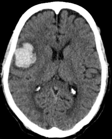
Figure 3: Image of the brain of a haemorrhage stroke patient [5]
Figure 3 illustrates the CT image of the brain of a haemorrhage stroke patient. There is a white area on the left side of the picture, which is where the bleeding occurs. Unlike ischemic stroke, haemorrhage stroke is less likely to onset. This is because vessel rupture is a rarer event compared to blood vessel blockage.
3. Application of AI in stroke treatment
With the development of technology, AI has gradually become an indispensable part of disease treatment. Due to AI's machine learning and deep learning, AI can often provide faster and more accurate diagnoses. Since stroke mainly causes damage to the brain, there are only two main ways to examine the brain: CT and MRI. They cannot present images well for judgment, so AI is particularly important in the diagnosis of stroke.
3.1. AI prediction of stroke
As a tool that can read various data in the brain, the brain-computer interface plays a big role in the prediction of stroke. Nowadays, the main brain-computer interfaces are divided into three types: Electroencephalography (EEG), Magnetoencephalography (MEG), and Electrocorticogram (ECoG). Among them, EEG is the most widely used. Brain-computer interfaces are divided into non-invasive and invasive. Among them, EEG and MEG are non-invasive. Although EEG is not as accurate as MEG, it is convenient and portable. For the EEG, there are three systems in total, (10-5), (10-10), and (10-20), and these three systems have different numbers of electrodes placed and the areas that can be detected. Among these systems, the most important one is the (10-20) system. The system is shown in Figure 4.
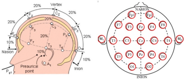
Figure 4: The (10-20) system of EEG [6]
EEG can detect and receive signals from the brain through electrodes, and then transmit them to a computer for AI to analyze these signals. Since machine learning requires samples, use EEG to sample and memorize brain information of stroke patients of different degrees and normal people. By using AI for machine learning, it can independently predict the possibility of a stroke [7]. Nowadays, AI can accomplish things that are difficult for humans to accomplish, such as image analysis. Due to the continuous increase in data, computing power is growing exponentially, and other advances. Machine learning and deep learning have made great improvements, such as the interpretation of images.
3.2. AI diagnosis of stroke
Nowadays, there are two common ways to examine the brain, CT and MRI. Compared with MRI, CT has gradually become the main way to examine stroke because of its low cost. Before the advent of thrombectomy, most stroke cases only required non-contrast CT as an evaluation requirement for suspected stroke patients. Additional imaging examinations are considered a waste of time and energy. Moreover, the non-contrast CT images of most stroke patients do not change much within 6 hours after the onset of symptoms, which means that non-contrast CT cannot diagnose stroke well. Moreover, when diagnosing ischemic stroke, the sensitivity of non-contrast CT for detecting acute ischemic tissue is only 26% [8].
AI can improve the speed of image generation as well as the quality. Some people oppose the use of advanced imaging techniques such as CT perfusion imaging or MRI because their acquisition and processing times tend to be longer than non-contrast CT and CT angiography. Most MRI stroke treatment protocols require scan times of less than 10 minutes, although MRI does require more detailed screening and patient transport time than CT. So improving the speed of image generation is necessary [9]. Generally, in the field of computer vision, Convolutional neural networks (CNNs) have been very successful in image classification of medical images.
3.3. AI rehabilitation of stroke
Since stroke is caused by the necrosis of some tissues in the brain, there will still be long-term or short-term sequelae after treatment, such as difficulty in movement, unclear speech, disorganization, cognitive errors, etc. Post-stroke rehabilitation is also very important. There are two main types of rehabilitation: Inpatient rehabilitation allows patients to receive appropriate rehabilitation training in the hospital. In contrast, the other type is Outpatient rehabilitation is training performed by the patient at home. Inpatient rehabilitation is good, and there are professional staff in the hospital to help patients recover. However, the gap between the number of stroke patients and the number of physicians is too large, which makes the work of physicians very busy.
Today, there is another way to automate recovery. Automated recovery systems can be divided into two main categories, robot-assisted and virtual reality-based. Robotic assistance mainly involves rehabilitation training with the help of exoskeletons or robots, while virtual reality mainly involves rehabilitation training in games [10]. With the addition of AI, this automated rehabilitation training has greatly improved the efficiency of rehabilitation, because AI can use deep learning and data obtained from exoskeletons or virtual reality to determine whether there is a better way.
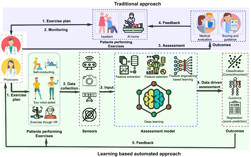
Figure 5: comparison of traditional and automated post-stroke rehabilitation [10]
Figure 5 shows the comparative overview between traditional and AI involved automated post-stroke rehabilitation. During the traditional approach, physicians need to take a lot of time to guide patients on how to do the exercise. In addition, it takes a lot of time for physicians to make the exercise plan, get feedback, and revise it. However, during the AI based automated approach, AI can replace physicians to analyze and make exercise plans. Through deep learning, the efficiency and accuracy of AI can help stroke patients conduct rehabilitation training more quickly and correctly.
4. Conclusions
As the population ages and stroke remains a major health concern, the integration of artificial intelligence and brain-machine interfaces offers a promising path forward in the fields of diagnosis, prediction, and rehabilitation. Ischemic and hemorrhagic strokes, although differing in cause, both result in significant brain damage that can severely impact the quality of life. Traditional diagnostic methods such as CT and MRI scans are valuable but limited in speed and sensitivity, particularly during the early stages of a stroke. AI, with its ability to analyze large datasets and recognize complex patterns, enhances the efficiency and accuracy of medical imaging, allowing for faster and more precise stroke diagnosis.
In rehabilitation, AI-driven systems using robotic assistance or virtual reality environments offer innovative solutions to bridge the gap between the demand for care and the availability of medical professionals. These systems can create personalized training plans and monitor patient progress in real time. Meanwhile, BMIs—especially non-invasive ones like EEG—serve as crucial tools for collecting brain activity data, enabling AI to predict stroke risk and assist in post-stroke recovery.
Despite these promising developments, several challenges remain. Invasive BMI systems pose risks and are costly, while non-invasive alternatives may lack precision. Additionally, the development of AI models requires vast amounts of quality data and ongoing refinement. Moving forward, interdisciplinary collaboration and further research will be essential to overcome these limitations. Ultimately, the fusion of AI and BMI holds significant potential to transform stroke care, offering hope for more effective treatment and improved patient outcomes.
References
[1]. Murphy, S. J., & Werring, D. J. (2020). Stroke: causes and clinical features. Medicine, 48(9), 561-566.
[2]. Xi, Z. (2024, November). Motion intention recognition based on AI and brain-machine interface. In IET Conference Proceedings CP901 (Vol. 2024, No. 24, pp. 305-309). Stevenage, UK: The Institution of Engineering and Technology.
[3]. Johns Hopkins Medicine. (2025, April 4). Brain anatomy and how the brain works. Retrieved from https://www.hopkinsmedicine.org/health/conditions-and-diseases/anatomy-of-the-brain
[4]. Lui, F., Hui, C., Khan, S. M. Z., et al. (2025, February 21). Ischemic stroke. In StatPearls [Internet]. Treasure Island, FL: StatPearls Publishing. Retrieved from https://www.ncbi.nlm.nih.gov/books/NBK499997/
[5]. Unnithan, A. K. A., Das, J. M., & Mehta, P. (2023, May 8). Hemorrhagic stroke. In StatPearls [Internet]. Treasure Island, FL: StatPearls Publishing. Retrieved from https://www.ncbi.nlm.nih.gov/books/NBK559173/
[6]. Orban, M., Elsamanty, M., Guo, K., Zhang, S., & Yang, H. (2022). A review of brain activity and EEG-based brain–computer interfaces for rehabilitation application. Bioengineering, 9(12), 768.
[7]. Islam, M. S., Hussain, I., Rahman, M. M., Park, S. J., & Hossain, M. A. (2022). Explainable artificial intelligence model for stroke prediction using EEG signal. Sensors, 22(24), 9859.
[8]. Bivard, A., Churilov, L., & Parsons, M. (2020). Artificial intelligence for decision support in acute stroke—current roles and potential. Nature Reviews Neurology, 16(10), 575-585.
[9]. Mouridsen, K., Thurner, P., & Zaharchuk, G. (2020). Artificial intelligence applications in stroke. Stroke, 51(8), 2573-2579.
[10]. Rahman, S., Sarker, S., Haque, A. N., Uttsha, M. M., Islam, M. F., & Deb, S. (2022). AI-driven stroke rehabilitation systems and assessment: A systematic review. IEEE Transactions on Neural Systems and Rehabilitation Engineering, 31, 192-207.
Cite this article
Xi,Z. (2025). The Tmpact of AI on Stroke Treatment and Prediction. Applied and Computational Engineering,166,13-19.
Data availability
The datasets used and/or analyzed during the current study will be available from the authors upon reasonable request.
Disclaimer/Publisher's Note
The statements, opinions and data contained in all publications are solely those of the individual author(s) and contributor(s) and not of EWA Publishing and/or the editor(s). EWA Publishing and/or the editor(s) disclaim responsibility for any injury to people or property resulting from any ideas, methods, instructions or products referred to in the content.
About volume
Volume title: Proceedings of CONF-SEML 2025 Symposium: Machine Learning Theory and Applications
© 2024 by the author(s). Licensee EWA Publishing, Oxford, UK. This article is an open access article distributed under the terms and
conditions of the Creative Commons Attribution (CC BY) license. Authors who
publish this series agree to the following terms:
1. Authors retain copyright and grant the series right of first publication with the work simultaneously licensed under a Creative Commons
Attribution License that allows others to share the work with an acknowledgment of the work's authorship and initial publication in this
series.
2. Authors are able to enter into separate, additional contractual arrangements for the non-exclusive distribution of the series's published
version of the work (e.g., post it to an institutional repository or publish it in a book), with an acknowledgment of its initial
publication in this series.
3. Authors are permitted and encouraged to post their work online (e.g., in institutional repositories or on their website) prior to and
during the submission process, as it can lead to productive exchanges, as well as earlier and greater citation of published work (See
Open access policy for details).
References
[1]. Murphy, S. J., & Werring, D. J. (2020). Stroke: causes and clinical features. Medicine, 48(9), 561-566.
[2]. Xi, Z. (2024, November). Motion intention recognition based on AI and brain-machine interface. In IET Conference Proceedings CP901 (Vol. 2024, No. 24, pp. 305-309). Stevenage, UK: The Institution of Engineering and Technology.
[3]. Johns Hopkins Medicine. (2025, April 4). Brain anatomy and how the brain works. Retrieved from https://www.hopkinsmedicine.org/health/conditions-and-diseases/anatomy-of-the-brain
[4]. Lui, F., Hui, C., Khan, S. M. Z., et al. (2025, February 21). Ischemic stroke. In StatPearls [Internet]. Treasure Island, FL: StatPearls Publishing. Retrieved from https://www.ncbi.nlm.nih.gov/books/NBK499997/
[5]. Unnithan, A. K. A., Das, J. M., & Mehta, P. (2023, May 8). Hemorrhagic stroke. In StatPearls [Internet]. Treasure Island, FL: StatPearls Publishing. Retrieved from https://www.ncbi.nlm.nih.gov/books/NBK559173/
[6]. Orban, M., Elsamanty, M., Guo, K., Zhang, S., & Yang, H. (2022). A review of brain activity and EEG-based brain–computer interfaces for rehabilitation application. Bioengineering, 9(12), 768.
[7]. Islam, M. S., Hussain, I., Rahman, M. M., Park, S. J., & Hossain, M. A. (2022). Explainable artificial intelligence model for stroke prediction using EEG signal. Sensors, 22(24), 9859.
[8]. Bivard, A., Churilov, L., & Parsons, M. (2020). Artificial intelligence for decision support in acute stroke—current roles and potential. Nature Reviews Neurology, 16(10), 575-585.
[9]. Mouridsen, K., Thurner, P., & Zaharchuk, G. (2020). Artificial intelligence applications in stroke. Stroke, 51(8), 2573-2579.
[10]. Rahman, S., Sarker, S., Haque, A. N., Uttsha, M. M., Islam, M. F., & Deb, S. (2022). AI-driven stroke rehabilitation systems and assessment: A systematic review. IEEE Transactions on Neural Systems and Rehabilitation Engineering, 31, 192-207.