


1. Introduction
With the rapid development of science and technology level and the continuous improvement of people's quality of life, the medical industry has developed greatly, but the contradiction between people's growing medical needs and the unbalanced development of medical resources has brought great challenges to the medical industry. Especially in the diagnosis of major diseases such as cancer, there is a higher demand for the accuracy and timeliness of the diagnostic results are increasingly demanding. Currently, clinical diagnosis of cancer mainly relies on imaging tests such as CT scans, PET imaging, MRI and X-ray [1]. Although these technologies can provide doctors with abundant diagnostic information, in the face of massive and complex medical imaging data, manual diagnosis is not only time-consuming and laborious, but also susceptible to factors such as doctors' fatigue, experience, and subjective judgment, leading to a decrease in diagnostic efficiency and accuracy. Therefore, there is an urgent need to introduce new technical means to improve diagnostic efficiency and accuracy.
In recent years, deep learning models have been widely used in the field of medical image analysis due to their excellent performance in processing large-scale data and image analysis. In particular, convolutional neural network (CNN) models have shown high potential in cancer diagnosis [2]. CNN models can automatically extract features from images in a short period of time and perform complex nonlinear combinations of these features, which can improve the accuracy and efficiency of cancer diagnosis.
This study compiles and summarizes the application of CNN models in cancer diagnosis over the past five years, focusing on the performance and progress of its application in different cancer types. It aims to provide valuable references for researchers related to this field and to promote the further application and optimization of CNN models in cancer diagnosis.
2. Application of CNN to cancer diagnosis
With the development of deep learning, CNN models have shown great value in medical image analysis and can be used for the diagnosis of many types of cancers, including brain tumors, lung cancer, breast cancer, liver cancer, skin cancer, etc. This automated diagnostic method can help doctors make diagnostic decisions more quickly and provide an effective means for early cancer diagnosis.
The excellent performance of the CNN model in medical imaging data processing is due to its structural characteristics, the structure of the CNN model is shown in figure1. As shown in the figure, the CNN model is mainly composed of convolutional layers, pooling layers, and activation functions. The utilization of convolutional layers effectively reduces the number of parameters by efficiently sharing weights, and meanwhile, in the training process, it automatically extracts the local features of the input data, which gives the CNN model good generalization ability. The pooling layer reduces the computational complexity of the network by decreasing the spatial size of the feature map while maintaining the most relevant features. Activation functions (e.g., ReLU) further improve the nonlinear representation of the model, making the CNN model perform better in processing complex medical images [3].
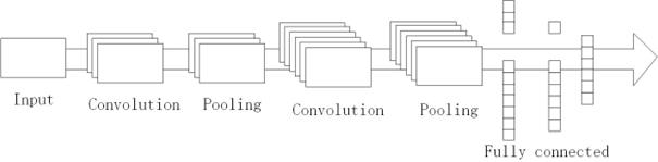
Figure 1. Structure of CNN model.
2.1. CNN model for lung cancer diagnosis
Lung cancer is characterized by a high incidence rate, high mortality rate and invisibility. In the early stage, lung cancer often has no obvious symptoms, and many patients have already entered the advanced stage by the time they are diagnosed, which increases the difficulty of treatment. As lung cancer grows fast, is prone to metastasis, and responds poorly to conventional treatments, early detection and diagnosis become especially important. Through early detection, patients can have more treatment options, thus improving treatment effects and survival rates [4]. To tackle the challenge of early diagnosis of lung cancer, 2DCNN was developed as a lightweight deep learning model for early diagnosis of lung cancer. The model employs the SMOTE algorithm to balance the dataset thus effectively tackling the problem of limited and unbalanced datasets, specifically, the use of the SMOTE algorithm to generate synthetic minority class samples can make the dataset more balanced in terms of the distribution of classes, thus improving the model performance. In terms of data processing, the 2DCNN model takes CT scan images as input and preprocesses the images. The steps include resizing the image to 256 × 256 pixels and applying a Gaussian filter for smoothing and noise reduction. Then the 2DCNN classifies the processed image. The model consists of three convolutional layers, three maximum pooling layers, one flat layer, and two fully connected layers, which are nonlinearly mapped using the ReLU activation function, and the CT scan images of the attendees are classified as normal, benign, or malignant using Softmax classifier. The dataset was split into training and test sets in an 8:2 ratio throughout the training phase. The model was trained using the Adam optimizer, with batch size set to 8 and training epoch set to 10. When the model was finally analyzed, it turned out to be 97% accurate [1].
2.2. CNN model for breast cancer diagnosis
Breast cancer is a serious cancer with an increasing incidence worldwide, partly because it is difficult to detect in its early stages. How breast cancer can be detected at an early stage and accurately treated and controlled is extremely important in reducing the mortality rate of breast cancer patients. Currently, commonly used methods in medical diagnosis include but are not limited to radiologic imaging, such as MRI, and mammography. However, these methods still have certain limitations, they all need to rely on doctors' clinical experience to make judgments, and the probability of misdiagnosis is high [5]. To address this problem, the pre-trained ResNet50v2 model is used to classify breast cancer images and optimized on the original basis. The positions of the batch normalization and activation functions were adjusted, the bottleneck structure was introduced, and global average pooling was performed on the last layer, which not only reduced the computational volume, but also effectively improved the training stability and convergence of the model.ResNet50v2 achieved a training accuracy of 80.17% and a validation accuracy of 81.70%.On the basis of ResNet50v2, the study also designed A 14-layer CNN model, which contains 1 input layer, 3 convolutional layers, 3 maximum pooling layers, 1 batch normalization layer, 1 global average pooling layer, 2 dropout layers, and 3 fully-connected layers, compared with the ResNet50v2 model, the 14-layer CNN model has an excellent performance, which is significantly better than the ResNet50v2 model. The ROC-AUC of the CNN model is calculated to be 0.82, and the confusion matrix of the model is given, both of which indicate that the model has a better performance on the classification task. The accuracy and loss epoch curves show a steady increase in accuracy and a decrease in the loss function during model training. [6]. In the diagnosis of breast cancer, a 20-layer feed-forward CNN model effectively denoises patchy noise in ultrasound images, followed by classification using a CNN model for accurate image classification. The 20-layer feed-forward CNN model estimates the original image by learning the noise features and performing residual mapping. Then another CNN classification model is used to classify the image which consists of the input layer, two hidden convolutional layers, a pooling layer, and an output layer. The method was evaluated on the Mendeley breast ultrasound dataset and the results showed that the method achieved excellent results on both classification tasks [7].
2.3. CNN model for brain tumor diagnosis
Brain tumors are one of the deadliest forms of cancer and they seriously affect the health of children and adults. Accurate diagnosis of the type and grade of brain tumors is particularly important in selecting appropriate treatment options. Until now, radiologists' manual examination of MRI images has been extremely time-consuming and error-prone. The emergence of automated computer-aided diagnosis (CAD) systems can well overcome these problems. Over the past few years, due to advances in artificial intelligence, CNN-based methods have been widely used for brain tumor classification and diagnosis, achieving good performance and results. However, the existing methods still have some limitations, such as insufficient classification accuracy, the need to manually segment the tumor region, and poor performance on small datasets [8]. The NawNet model is based on the AlexNet model, and the model is further optimized by adding different types of layers, adjusting the size of the filter, and using dropout layers. Nine convolutional blocks, two fully connected layers, and one output layer make up the model's total of 22 layers of low complexity architecture. Each convolutional block has a convolutional layer, a ReLU activation layer, and a batch normalization layer. The fully connected layer uses a dropout layer to prevent overfitting, and the final convolutional layer uses an average pooling layer to improve model accuracy. The model was trained on Kaggle's public dataset which contains 7022 MRI images, from which 3350 images were selected for experiments to get 99.4% accuracy of NawNet model [9]. Another study pointed out that such as EfficientNetB0, MobileNetV2 and Xception by optimizing the original CNN model architecture and then classifying the brain tumor images from MRI scans. Firstly, based on the original structure, the performance of the model is improved by introducing an additional pooling layer, dropout layer and ReLu activation function. The training process of the deep learning model is accelerated by introducing the concept of migration learning, which directly reads the pre-trained weights from the ImageNet dataset. Pre-processing of brain MRI image data, including adjusting resolution, horizontal rotation of images and other operations to improve the quality and quantity of the dataset. To guarantee the dataset's unpredictability, the brain tumor photos were split into training, test, and validation sets during the training procedure. Ultimately, the accuracy of the CNN variant model mentioned above is computed [10].
3. Results and discussion
In this article, the performance of different CNN models in the diagnosis of lung cancer, breast cancer and brain tumors is organized and the accuracy of each model is compared and analyzed, and the specific results are shown in table1.
Table 1. Comparison of results of different CNN models in cancer diagnosis.
Model |
Dataset |
Type of cancer |
Accuracy |
2DCNN [1] |
Iraq Oncology Teaching Hospital/National Cancer Disease Center |
Lung Cancer |
97% |
ResNet50v2 [6] |
BreaKHis |
Breast Cancer |
80.17% |
CNN Denoising and Classification model [7] |
Mendeley |
Breast Cancer |
88% |
NawNet [9] |
Kaggle |
Brain Tumor |
99.4% |
MobileNetV2 [10] |
ImageNet |
Brain Tumor |
98.80% |
EfficientNetB0 [10] |
ImageNet |
Brain Tumor |
98% |
Xception [10] |
ImageNet |
Brain Tumor |
97% |
Table 1 shows the performance of three different CNN models in the diagnosis of lung cancer, breast cancer and brain tumor. It can be seen that in the diagnosis of lung cancer, the CNN model reduces the cost of manual diagnosis by medical personnel and improves the diagnostic efficiency of lung cancer, but a large amount of data about lung cancer is needed in the future for the training of the model, and the use of deep learning models for diagnosis inevitably involves the annotation of the dataset and the patient's privacy [1]. Meanwhile, the 2DCNN model combined with the SMOTE algorithm proposed in this study achieved an accuracy of 97%, but the method is still limited and cannot be applied to all datasets.
In research on breast cancer, the CNN model has performed well in assisting with early breast cancer detection and helping to reduce the lethality of the disease. For future research, the proposed 14-layer CNN model can be further optimized and improved to increase the performance and accuracy of the model. Also consider how the model can be applied to more fields, such as skin cancer, lung cancer, etc. To increase the model's accuracy, further study should be done on the extraction of valid feature values from healthcare impacts and use them as model training data.
In the CNN model for diagnosis of brain tumors, the NawNet model has less complexity but high accuracy compared to existing methods. This CNN model is currently trained using only a limited dataset, and in the future, we can try to continue to expand the dataset or explore some data enhancement techniques to further improve the classification performance of this model. Currently, the model only uses the CNN model, other more advanced models can be considered in the future to further optimize the structure and improve the accuracy of the classification task. In summary, generalization and extension of the CNN model is particularly important in clinical medicine, as well as for other more advanced models (e.g., Transformer, etc.). Optimizing the model structure, collecting larger datasets, and training the model better are directions for future research to be done.
CNN models show a large potential for application in clinical cancer tumor diagnosis. However, the performance and speed of CNN models, in terms of the quantity and quality of datasets, are still key directions for future research. In the future, more sophisticated models may be employed to increase classification task accuracy and offer more precise and trustworthy medical diagnosis instruments.
4. Conclusion
Cancer, as a serious disease, poses a huge health threat and economic burden to human society. In the traditional medical diagnosis process, medical personnel mainly rely on manual methods to diagnose various cancers. This diagnostic method not only consumes a lot of time and increases medical costs, but also relies on the personal experience of medical personnel, resulting in poor cancer treatment results. To improve this situation, the introduction of computerized deep learning technology for assisted diagnosis has become an important development direction in the medical field. This technology can assist medical personnel in classifying features such as medical images, thereby improving the early diagnosis rate of various cancers to facilitate further examination in the future. In recent years, classification techniques for different cancers have been rapidly developed, among which CNN models have achieved remarkable results in the field of medical diagnosis.
The structural features of CNN models enable them to exhibit excellent performance in image classification tasks. However, there are still some limitations of CNN models in practical applications. The main issues that remain to be solved include developing novel model architectures to enhance model performance, expanding the training dataset to overcome the dilemma of insufficient data volume for diagnostic accuracy, and enhancing the generalization ability of the model so that it can better adapt to different types of cancer diagnosis. Solving the above problems will have a profound impact on the work of healthcare professionals and greatly reduce their workload, thus driving a major breakthrough in human health management. On this basis, we are expected to realize more efficient and accurate cancer diagnosis, provide better medical services to patients, and ultimately improve the overall cure rate of cancer.
References
[1]. Begum S H, Baig M I, Hussain M A and Muqeet M A. (2022) A Lightweight Deep Learning Model for Automatic Diagnosis of Lung Cancer. 2022 IEEE 2nd International Conference on Mobile Networks and Wireless Communications (ICMNWC), Tumkur, Karnataka, India, pp. 1-5
[2]. Gasmi M, Derdour M, Gahmousse A, Amroune M, Bendjenna H and Sahraoui B. (2021) Multi-Input CNN for molecular classification in breast cancer. 2021 International Conference on Recent Advances in Mathematics and Informatics (ICRAMI), Tebessa, Algeria pp. 1-5
[3]. [Dai D. An Introduction of CNN: Models and Training on Neural Network Models. (2021) 2021 International Conference on Big Data, Artificial Intelligence and Risk Management (ICBAR), Shanghai, China, pp. 135-138
[4]. Makandar A and Jadhav M N. Disease Recognition in Medical Images Using CNN-LSTM-GRU Ensemble, a Hybrid Deep Learning. (2023) 2023 7th International Conference on Computation System and Information Technology for Sustainable Solutions (CSITSS), Bangalore, India, pp. 1-9
[5]. Gasmi M, Derdour M, Gahmousse A, Amroune M, Bendjenna H and Sahraoui B. Multi-Input CNN for molecular classification in breast cancer. (2021) 2021 International Conference on Recent Advances in Mathematics and Informatics (ICRAMI), Tebessa, Algeria, pp. 1-5
[6]. Sharma G, Anand V and Gupta S. Ensembling of Deep Learning Models for Automatic Diagnosis of Breast Cancer with Histopathology Images. (2023) 2023 Global Conference on Information Technologies and Communications (GCITC), Bangalore, India, pp. 1-6
[7]. Latif G, Butt M O, Yousif Al Anezi F and Alghazo J. Ultrasound Image Despeckling and detection of Breast Cancer using Deep CNN. (2020) 2020 RIVF International Conference on Computing and Communication Technologies (RIVF), Ho Chi Minh City, Vietnam, pp. 1-5
[8]. Ayadi W, Elhamzi W and Atri M. A new deep CNN for brain tumor classification. (2020) 2020 20th International Conference on Sciences and Techniques of Automatic Control and Computer Engineering (STA), Monastir, Tunisia, pp. 266-270
[9]. Al-Ani N and Al-Shamma O. Implementing a Novel Low Complexity CNN Model for Brain Tumor Detection. (2020) 2022 8th International Conference on Contemporary Information Technology and Mathematics (ICCITM), Mosul, Iraq, pp. 358-363
[10]. Indra Z, Jusman Y and Kurniawan R. Development of Deep Learning Model Base on Modified CNN Architectures for Brain Tumours Early Diagnosis. (2023) 2023 3rd International Conference on Electronic and Electrical Engineering and Intelligent System (ICE3IS), Yogyakarta, Indonesia, pp. 503-507
Cite this article
Li,X. (2024). CNN-Based Cancer Image Diagnosis: Current Progress and Future Directions. Applied and Computational Engineering,106,7-12.
Data availability
The datasets used and/or analyzed during the current study will be available from the authors upon reasonable request.
Disclaimer/Publisher's Note
The statements, opinions and data contained in all publications are solely those of the individual author(s) and contributor(s) and not of EWA Publishing and/or the editor(s). EWA Publishing and/or the editor(s) disclaim responsibility for any injury to people or property resulting from any ideas, methods, instructions or products referred to in the content.
About volume
Volume title: Proceedings of the 2nd International Conference on Machine Learning and Automation
© 2024 by the author(s). Licensee EWA Publishing, Oxford, UK. This article is an open access article distributed under the terms and
conditions of the Creative Commons Attribution (CC BY) license. Authors who
publish this series agree to the following terms:
1. Authors retain copyright and grant the series right of first publication with the work simultaneously licensed under a Creative Commons
Attribution License that allows others to share the work with an acknowledgment of the work's authorship and initial publication in this
series.
2. Authors are able to enter into separate, additional contractual arrangements for the non-exclusive distribution of the series's published
version of the work (e.g., post it to an institutional repository or publish it in a book), with an acknowledgment of its initial
publication in this series.
3. Authors are permitted and encouraged to post their work online (e.g., in institutional repositories or on their website) prior to and
during the submission process, as it can lead to productive exchanges, as well as earlier and greater citation of published work (See
Open access policy for details).
References
[1]. Begum S H, Baig M I, Hussain M A and Muqeet M A. (2022) A Lightweight Deep Learning Model for Automatic Diagnosis of Lung Cancer. 2022 IEEE 2nd International Conference on Mobile Networks and Wireless Communications (ICMNWC), Tumkur, Karnataka, India, pp. 1-5
[2]. Gasmi M, Derdour M, Gahmousse A, Amroune M, Bendjenna H and Sahraoui B. (2021) Multi-Input CNN for molecular classification in breast cancer. 2021 International Conference on Recent Advances in Mathematics and Informatics (ICRAMI), Tebessa, Algeria pp. 1-5
[3]. [Dai D. An Introduction of CNN: Models and Training on Neural Network Models. (2021) 2021 International Conference on Big Data, Artificial Intelligence and Risk Management (ICBAR), Shanghai, China, pp. 135-138
[4]. Makandar A and Jadhav M N. Disease Recognition in Medical Images Using CNN-LSTM-GRU Ensemble, a Hybrid Deep Learning. (2023) 2023 7th International Conference on Computation System and Information Technology for Sustainable Solutions (CSITSS), Bangalore, India, pp. 1-9
[5]. Gasmi M, Derdour M, Gahmousse A, Amroune M, Bendjenna H and Sahraoui B. Multi-Input CNN for molecular classification in breast cancer. (2021) 2021 International Conference on Recent Advances in Mathematics and Informatics (ICRAMI), Tebessa, Algeria, pp. 1-5
[6]. Sharma G, Anand V and Gupta S. Ensembling of Deep Learning Models for Automatic Diagnosis of Breast Cancer with Histopathology Images. (2023) 2023 Global Conference on Information Technologies and Communications (GCITC), Bangalore, India, pp. 1-6
[7]. Latif G, Butt M O, Yousif Al Anezi F and Alghazo J. Ultrasound Image Despeckling and detection of Breast Cancer using Deep CNN. (2020) 2020 RIVF International Conference on Computing and Communication Technologies (RIVF), Ho Chi Minh City, Vietnam, pp. 1-5
[8]. Ayadi W, Elhamzi W and Atri M. A new deep CNN for brain tumor classification. (2020) 2020 20th International Conference on Sciences and Techniques of Automatic Control and Computer Engineering (STA), Monastir, Tunisia, pp. 266-270
[9]. Al-Ani N and Al-Shamma O. Implementing a Novel Low Complexity CNN Model for Brain Tumor Detection. (2020) 2022 8th International Conference on Contemporary Information Technology and Mathematics (ICCITM), Mosul, Iraq, pp. 358-363
[10]. Indra Z, Jusman Y and Kurniawan R. Development of Deep Learning Model Base on Modified CNN Architectures for Brain Tumours Early Diagnosis. (2023) 2023 3rd International Conference on Electronic and Electrical Engineering and Intelligent System (ICE3IS), Yogyakarta, Indonesia, pp. 503-507