


1. Introduction
Nanomaterials refer to materials that have at least one dimension in the nanometer range (1-100nm) or are composed of such dimensions as basic units. This microscopic scale gives them vastly different physical, chemical and biological properties from macroscopic materials, providing an unprecedented opportunity to solve long-standing problems in the medical field. By virtue of their special size, nanomaterials exhibit three major effects that distinguish them from traditional materials: small-size effects, surface effects and quantum-size effects.
These unique effects give nanomaterials many excellent properties. In terms of mechanical properties, the strength and hardness of nano-metal materials are much higher than those of traditional metals. Regarding optical properties, nanomaterials have special optical absorption and emission characteristics, which can be used to manufacture efficient optoelectronic devices. In terms of chemical properties, their high specific surface area endows nanomaterials with extremely high catalytic activity, leading to broad application prospects in chemical industry, environmental protection and other fields.
Nowadays, nanomaterials have been widely used in many fields. In the rapidly advancing landscape of modern medical science and technology, nanomaterials, with their unique properties and diverse functions, have gradually emerged as a key force promoting continuous progress in the medical field. In the medical field, nanoparticles can be used as drug carriers to achieve precise drug delivery and improve treatment efficiency and effect. This paper focuses on the mechanism of action and application prospect of several kinds of nanomaterials in drug delivery, medical diagnosis and treatment, offering insights that may guide future research directions and practical uses of nanomaterials in the medical field.
2. Nanomaterials Commonly Used in Drug Delivery
Several types of nanomaterials are described below for use in several areas of medical care.
2.1. Magnetic Nanoparticles
In terms of targeted drug delivery, as a nanoparticle, it shows significant advantages. Its large specific surface area enables it to absorb proteins or carry drugs, providing a solid foundation for targeted drug delivery. Additionally, magnetic nanoparticles possess responsiveness to external magnetic fields, enabling researchers to easily separate them from mixture systems using magnets. This characteristic makes them widely applicable in separation techniques, such as cell separation.
Second, magnetic nanoparticles are superparamagnetic, which allows them to concentrate precisely at the desired site when an external magnetic field is applied. In the biomedical field, the most commonly used magnetic nanoparticles are Fe, Co, Ti, Ni alloys and iron oxide, ferrites (such as BaFeO12 and CoFe2O4). Among them, iron oxide nanoparticles (usually Fe2O3 or FeO) have been extensively studied due to their low toxicity and high biocompatibility [1].
In the late 1970s, Widder et al. proposed the idea of placing high magnetic fields at tumor sites and then using magnetic nanoparticles to carry drugs to specific sites [2]. Subsequent research demonstrated the effectiveness of this approach in numerous small animal studies, and even a small number of clinical trials. Figure 1 illustrates the preparation and drug loading mechanism of porous, hollow magnetic nanomaterials.
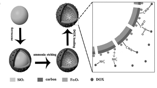
Figure 1: Schematic diagram of preparation and drug loading of porous hollow Fe3O4@C nanocapsules [2]
Magnetic nanomaterials also perform well in controlled drug release. The rate and time of drug release from the nanocarriers can be controlled by using the thermal effect or mechanical vibration generated by the alternating magnetic field. For example, during a specific treatment period, researchers can adjust the magnetic field parameters to prompt the nanocarrier to release the drug on demand, thereby maintaining a stable and effective drug concentration. This mechanism not only improves the drug's efficacy, but also reduces side effects, leading to a significant improvement in the patient's treatment experience. As research advances, it is expected that more innovative applications based on magnetic nanoparticles will be developed in the future, providing more effective tools for clinical treatment.
The template method for the synthesis of magnetic micro-nanostructures has a long history and is also one of the most commonly used method for the synthesis of magnetic hollow nanoparticles. For example, Zhao et al reported a method for preparing hollow mesoporous silica spheres with a particle size of about 100 nm by micellar aggregate template method [3]. Cao et al have successfully developed a new two-step precursor template conversion method to prepare layered nanostructured magnetic hollow spheres assembled with Fe3O4 or gamma-Fe2O3 nanosheets [4]. Figure 2 shows the preparation process of hollow magnetic nanoparticles.
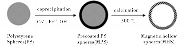
Figure 2: Preparation process of hollow magnetic nanoparticles [4]
2.2. Ethylene Glycol Targeting Nanocarriers
Polyethylene glycol (PEG) has gained widespread use in the design of targeted nanocrimers due to its good hydrophilicity, biocompatibility and low immunogenicity, significantly driving the field of drug delivery.
Peg-modified nanocarriers can extend their time in blood circulation. Under normal physiological conditions, nanocarriers are easily recognized and cleared by the immune system. However, the hydrophilic shell of PEG reduces protein adsorption and macrophage uptake, thereby significantly prolonging their half-life [5]. Currently approved nanomedical drugs include Doxil (a PEG-coated liposome nanomedical drug containing doxorubicin), used for treating cancers such as breast cancer, ovarian cancer and Kaposi sarcoma; Abraxane (a nanoparticle protein binding containing paclitaxel), used for breast cancer, non-small cell lung cancer and pancreatic cancer; and Onivyde (a PEG-coated liposome nanocide containing irinotecan), which is indicated for treat pancreatic cancer [6]. Each of these drugs has demonstrated clinical success.
In terms of enhancing targeting, PEG can be used as a connecting arm to connect targeted molecules (such as antibodies, peptides, aptamers) to the surface of nanocarriers to achieve precise delivery to specific tissues or cells. Taking PEG-modified nanoparticles connected to tumor-specific antibodies as an example, it can effectively increase the drug concentration at the tumor site and enhance the anti-tumor effect.
According to the current research progress, nanomaterials have broad application prospects in drug delivery. The nanomaterial drug delivery system can achieve precise targeted delivery to tumor tissue. By modifying the surface with specific targeting ligands, nanocarriers can recognize and bind to specific receptors on the surface of tumor cells, efficiently delivering drugs to the tumor site while reducing the toxic side effects on normal tissues, and improving the therapeutic effect. In addition, nanomaterials can also function as gene carriers, safely and effectively delivering therapeutic genes to target cells, thus providing a new way for gene therapy.
3. Multifunctional Nanoprobes: Commonly Used Nanomaterials in Medical Diagnostic Devices
In the early stages of cancer, trace amounts of tumor markers appear in the body. With its high sensitivity and specificity, multifunctional bionanometers can accurately identify and detect these markers. For example, gold nanoparticles (AuNPs) have become a research hotspot in the field of tumor diagnosis and treatment due to their excellent optical and thermodynamic properties, biocompatibility and easy surface functionalization [7-8]. The nanoprobes made from AuNPs have excellent optical properties and X-ray attenuation ability, which can be used in fluorescence imaging and computed tomography (CT) imaging [9]. In addition to having good imaging effects, AuNPs also demonstrate effective photothermal therapy (PTT) effects [10-11]. AuNPs can convert the absorbed near-infrared light (NIR) into heat, increasing the temperature within tumor cells, promoting cell apoptosis, and eventually leading to thermal ablation of tumor [12-13]. This detection method allows for the identification of abnormalities in the very early stages of cancer, significantly improving the rate of early diagnosis of cancer.
In the prevention and control of infectious diseases, rapid and accurate detection of pathogens is crucial. Bio-nano probes labeled with quantum dots perform well in this regard. Quantum dots have unique fluorescence characteristics, high stability and high fluorescence intensity. By combining them with specific nucleic acids or proteins that recognize pathogens, highly sensitive probes for pathogen detection can be constructed. In the detection process, once the probe binds to the pathogen, the presence and type of the pathogen can be quickly determined through the change of the fluorescent signal, greatly shortening the detection time.
With the continuous development of nanotechnology and biomedicine, nano-antibacterial biomaterials are expected to have a broader application prospect in the field of medical treatment. By optimizing the preparation process of nanomaterials, researchers can improve their biosafety and stability. In addition, strengthening the cross-fusion of nano-antibacterial biomaterials with other technologies will help develop more innovative medical products. Finally, the mechanism of action and metabolic pathway of nano-antibacterial biomaterials in vivo is necessary to provide a more solid theoretical foundation for their clinical application.
4. Nanomaterials Commonly Used in Therapeutic Methods
Nano silver possesses properties such as surface-enhanced Raman scattering, making it a promising substrate for improving detection sensitivity in cancer diagnosis.
Silver nanoparticles enter cancer cells mainly through endocytosis and can also cross the blood-brain barrier due to their tiny size. They exhibit cytotoxic effects on cancer cells by reducing mitochondrial function, generating reactive oxygen species, releasing lactate dehydrogenase, inducing cell cycle dysregulation, and up-regulating apoptosis-related genes like Bax. Additionally, they can cause micronucleus formation, chromosomal aberrations, and DNA damage. Morphologically, their effects on cells include rupturing, aggregation, contraction, non-adherence, and loss of membrane stability.
Researchers have conducted a large number of experiments on lung cancer cells. Pan Xiaofang et al. found that after nano silver was applied to lung cancer A549 cells for 48 hours, some cells began to exhibit blurred and wrinkled outlines, with a decrease in volume, leading to irregular shapes [14]. With the increase of nano silver concentration, so did the number of apoptotic A549 cells. In vitro experiments confirmed that silver nanoparticles could arrest the cell cycle of A549 cells in G2/M phase, disorder the expression of P21 and P53 genes and induce the expression of apoptosis factors, thus inhibiting the growth of cancer cells through multi-target effects. Similarly, BLANCO et al. found that in lung cancer A549 cells, daily low dose of nano silver or single high dose of nano silver exposure for 72h could reduce the expression of genes and proteins such as P21, P53, MDM2 and caspase-3, while increasing the generation of reactive oxygen species and cause DNA damage. This ultimately led to the inhibition of cancer cell proliferation. As shown in Figure 3, various mechanisms of apoptosis induced by silver nanoparticles in cancer cells are depicted.
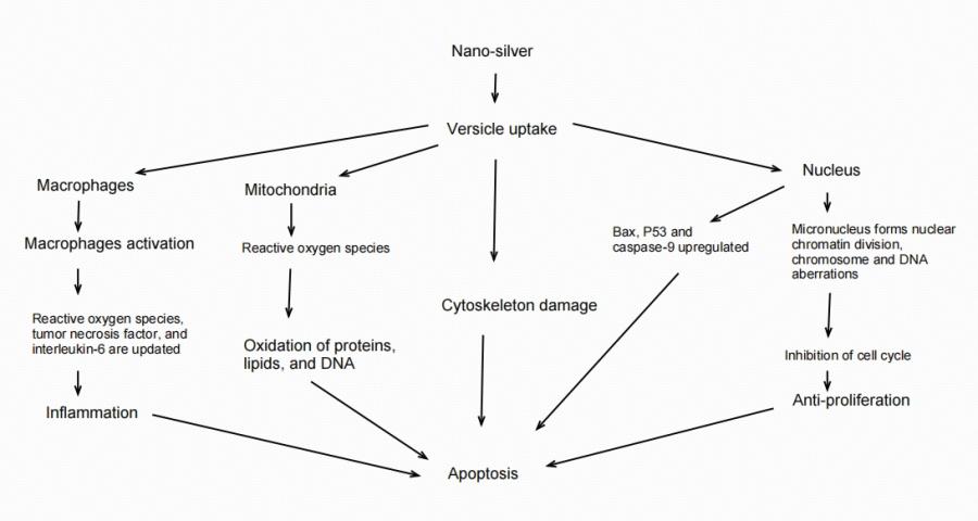
Figure 3: Anti-cancer mechanism of nano-silver [14]
5. Existing Challenges
As an emerging material, nanomaterials have made remarkable progress in applied research in the medical field due to their unique small-size effects, surface effects and quantum size effects, providing new ideas and methods for solving medical problems and promoting medical progress. However, the nanomedicine industry still faces many challenges from basic research to the development of large-scale medical products.
Firstly, biosafety is a significant concern. There is limited understanding of the long-term effects and potential toxicity of nanomaterials once they enter the human body. Some nanomaterials may accumulate in specific organs or tissues in the body, leading to inflammatory reactions, oxidative stress, genotoxicity and other adverse consequences.
Secondly, the mechanism of action of nanomaterials in living organisms is still unclear. Although nanomaterials have shown good performance in vitro experiments, their behavior and mode of action may change significantly in the complex physiological environment in vivo. After entering the human body, nanomaterials will interact with proteins, cells and other components in the blood, which will change the surface properties and biological activities of nanomaterials, which can affect their distribution, metabolism and targeting in the body.
In addition, large-scale preparation and quality control are also urgent problems to be solved. Some preparation methods require extreme conditions—such as high temperature, high pressure, or high vacuum—making large-scale production difficult and costly. Moreover, quality control is a serious issue. The properties of nanomaterials are highly sensitive to factors such as their size, shape and surface chemistry, and even small changes in preparation conditions can lead to significant differences in product quality.
Finally, the difficulties of clinical transformation should not be ignored. Developing nanomaterials demands substantial funding for basic research, clinical trials, and production facilities. Strict regulatory requirements have also brought great challenges to the clinical transformation of nanomaterials. Regulatory authorities have strict standards for the safety, effectiveness and quality controllability of nanomaterials. Therefore, nanomaterials need to go through a large number of clinical trials to prove their safety and effectiveness, a process that is time-consuming and labor-intensive.
Despite their broad application potential in the medical field, nanomaterials still face many challenges in their development. Further research and innovation are needed to overcome these obstacles.
6. Conclusion
This paper explores the application of nanomaterials in the medical field, covering multiple aspects of drug delivery, medical diagnosis, and treatment methods. The action mechanism and application prospect of several typical nanomaterials are also introduced in this paper. Studies have shown that nanomaterials can effectively improve the therapeutic efficiency of drugs in cancer and other diseases by virtue of their characteristics of high stability, high specificity and high sensitivity. However, there are some shortcomings in this paper, including a lack of concrete empirical data, insufficient discussion on the long-term biosafety and mechanisms of action of nanomaterials, and a lack of systematic evaluation of existing studies.
Future research should focus on several key areas. A deeper understanding of how nanomaterials interact within the human body is essential. It is also crucial to identify the toxicity and side effects of nanomaterials on the human body, and reduce the possible toxicity and side effects in the human body. Although nanomaterials have broad prospects in the medical field, the problems they face cannot be ignored. Addressing these issues will maximize the benefits of nanomaterials, ultimately contributing to improved health outcomes and well-being for patients.
References
[1]. Wang, L., Liu, L., Jin, X., et al. (2021). Preparation of magnetic nanoparticles and their application in targeted drug delivery. Guangzhou Chemical Industry, 49(14), 14-17.
[2]. Wang, D. J., Zhang, J. Y., He, P., & et al. (2019). Size-modulated electromagnetic properties and highly efficient microwave absorption of magnetic iron oxide ceramic opened-hollow microspheres. Ceramics International, 45(17), 23043-23049.
[3]. Zhao, W., Lang, M., Li, Y., & et al. (2009). Fabrication of uniform hollow mesoporous silica spheres and ellipsoids of tunable size through a facile hard-templating route. Journal of Materials Chemistry, 19, 2778-2783.
[4]. Cao, S. W., Zhu, Y. J., Ma, M. Y., & et al. (2008). Hierarchically nanostructured magnetic hollow spheres of Fe₃O₄ and γ-Fe₂O₃: Preparation and potential application in drug delivery. Journal of Physical Chemistry C, 112(6), 1851-1856.
[5]. Wang, J. (2018). Control of PEGylation degree on nanocarrier surfaces and its research in drug delivery [D]. Hefei: University of Science and Technology of China.
[6]. Li Y Nan. (2023). Study on the mechanism of action and anti-tumor effect of bionomanite drug assembly targeting and enhancing oxidative stress in breast cancer [D]. Chongqing: Chongqing University.
[7]. Fan, M., Han, Y., Gao, S. T., et al. (2020). Ultrasmall gold nanoparticles in cancer diagnosis and therapy. Theranostics, 10(11), 4944-4957.
[8]. Chen, Y., Montana, D. M., Wei, H., & et al. (2017). Shortwave infrared in vivo imaging with gold nanoclusters. Nano Letters, 17(10), 6330-6334.
[9]. Wu, Y., Ali, M. R. K., Chen, K. C., et al. (2019). Gold nanoparticles in biological optical imaging. Nano Today, 24, 120-140.
[10]. Mao, Q. L., Fang, J., Wang, A. N., et al. (2021). Aggregation of gold nanoparticles triggered by hydrogen peroxide-initiated chemiluminescence for activated tumor theranostics. Angewandte Chemie International Edition, 60(44), 23805-23811.
[11]. Li, H., Wang, P., Deng, Y. X., & et al. (2017). Combination of active targeting, enzyme-triggered release and fluorescent dye into gold nanoclusters for endomicroscopy-guided photothermal/photodynamic therapy to pancreatic ductal adenocarcinoma. Biomaterials, 139, 30-38.
[12]. Jain, P. K., Huang, X. H., El-Sayed, I. H., et al. (2008). Noble metals on the nanoscale: Optical and photothermal properties and some applications in imaging, sensing, biology, and medicine. Accounts of Chemical Research, 41(12), 1578-1586.
[13]. Cheng, L., Wang, C., Feng, L. Z., & et al. (2014). Functional nanomaterials for phototherapies of cancer. Chemical Reviews, 114(21), 10869-10939.
[14]. Liu, R., Xu, J., Hu, J., et al. (2019). The application of nano-silver in cancer diagnosis and treatment. Chinese Journal of Advanced Medicine & Clinic, 39(05), 268-273.
Cite this article
Zhao,G. (2025). Application of Nanomaterials in Medical Field. Applied and Computational Engineering,142,221-226.
Data availability
The datasets used and/or analyzed during the current study will be available from the authors upon reasonable request.
Disclaimer/Publisher's Note
The statements, opinions and data contained in all publications are solely those of the individual author(s) and contributor(s) and not of EWA Publishing and/or the editor(s). EWA Publishing and/or the editor(s) disclaim responsibility for any injury to people or property resulting from any ideas, methods, instructions or products referred to in the content.
About volume
Volume title: Proceedings of MSS 2025 Symposium: Automation and Smart Technologies in Petroleum Engineering
© 2024 by the author(s). Licensee EWA Publishing, Oxford, UK. This article is an open access article distributed under the terms and
conditions of the Creative Commons Attribution (CC BY) license. Authors who
publish this series agree to the following terms:
1. Authors retain copyright and grant the series right of first publication with the work simultaneously licensed under a Creative Commons
Attribution License that allows others to share the work with an acknowledgment of the work's authorship and initial publication in this
series.
2. Authors are able to enter into separate, additional contractual arrangements for the non-exclusive distribution of the series's published
version of the work (e.g., post it to an institutional repository or publish it in a book), with an acknowledgment of its initial
publication in this series.
3. Authors are permitted and encouraged to post their work online (e.g., in institutional repositories or on their website) prior to and
during the submission process, as it can lead to productive exchanges, as well as earlier and greater citation of published work (See
Open access policy for details).
References
[1]. Wang, L., Liu, L., Jin, X., et al. (2021). Preparation of magnetic nanoparticles and their application in targeted drug delivery. Guangzhou Chemical Industry, 49(14), 14-17.
[2]. Wang, D. J., Zhang, J. Y., He, P., & et al. (2019). Size-modulated electromagnetic properties and highly efficient microwave absorption of magnetic iron oxide ceramic opened-hollow microspheres. Ceramics International, 45(17), 23043-23049.
[3]. Zhao, W., Lang, M., Li, Y., & et al. (2009). Fabrication of uniform hollow mesoporous silica spheres and ellipsoids of tunable size through a facile hard-templating route. Journal of Materials Chemistry, 19, 2778-2783.
[4]. Cao, S. W., Zhu, Y. J., Ma, M. Y., & et al. (2008). Hierarchically nanostructured magnetic hollow spheres of Fe₃O₄ and γ-Fe₂O₃: Preparation and potential application in drug delivery. Journal of Physical Chemistry C, 112(6), 1851-1856.
[5]. Wang, J. (2018). Control of PEGylation degree on nanocarrier surfaces and its research in drug delivery [D]. Hefei: University of Science and Technology of China.
[6]. Li Y Nan. (2023). Study on the mechanism of action and anti-tumor effect of bionomanite drug assembly targeting and enhancing oxidative stress in breast cancer [D]. Chongqing: Chongqing University.
[7]. Fan, M., Han, Y., Gao, S. T., et al. (2020). Ultrasmall gold nanoparticles in cancer diagnosis and therapy. Theranostics, 10(11), 4944-4957.
[8]. Chen, Y., Montana, D. M., Wei, H., & et al. (2017). Shortwave infrared in vivo imaging with gold nanoclusters. Nano Letters, 17(10), 6330-6334.
[9]. Wu, Y., Ali, M. R. K., Chen, K. C., et al. (2019). Gold nanoparticles in biological optical imaging. Nano Today, 24, 120-140.
[10]. Mao, Q. L., Fang, J., Wang, A. N., et al. (2021). Aggregation of gold nanoparticles triggered by hydrogen peroxide-initiated chemiluminescence for activated tumor theranostics. Angewandte Chemie International Edition, 60(44), 23805-23811.
[11]. Li, H., Wang, P., Deng, Y. X., & et al. (2017). Combination of active targeting, enzyme-triggered release and fluorescent dye into gold nanoclusters for endomicroscopy-guided photothermal/photodynamic therapy to pancreatic ductal adenocarcinoma. Biomaterials, 139, 30-38.
[12]. Jain, P. K., Huang, X. H., El-Sayed, I. H., et al. (2008). Noble metals on the nanoscale: Optical and photothermal properties and some applications in imaging, sensing, biology, and medicine. Accounts of Chemical Research, 41(12), 1578-1586.
[13]. Cheng, L., Wang, C., Feng, L. Z., & et al. (2014). Functional nanomaterials for phototherapies of cancer. Chemical Reviews, 114(21), 10869-10939.
[14]. Liu, R., Xu, J., Hu, J., et al. (2019). The application of nano-silver in cancer diagnosis and treatment. Chinese Journal of Advanced Medicine & Clinic, 39(05), 268-273.