1. Introduction
A tumor refers to a cluster of cells that exhibits abnormal proliferation and growth, resulting from the failure of regulatory mechanisms accountable for normal cell cycle progression. Brain tumors are classified into two; low grade (grades 1 and 2), and high grade (grades 3 and 4), where the former is benign, and the latter is malignant. The benign are non-cancerous and do not spread, while the malignant are cancerous and spread indefinitely. A brain tumor is a fatal type of cancer affecting children and adults. Brain tumors are of various types depending on the area affected, whether benign or malignant. It has been found to have a low survival rate due to a high level of misdiagnosis, leading to poor treatment choices. Incorrect classification of brain tumors is a significant challenge that leads to undesirable prognosis, diagnosis, and treatment consequences [1]. Correct and early diagnosis and treatment is one factor that leads to an increased survival rate for patients with brain tumors.
According to the World Health Organization, people suffer from brain tumors are appropriately 700,000. As of 2019, the number of people diagnosed with brain tumors is 86,000, and those who have died of brain tumors are 16,830—an overall survival rate of 35% [2]. Manual extraction of brain tumor features and categorization is prone to a high level of errors and also time-consuming; therefore, an automated approach to brain tumor diagnosis is needed. The authors in [3] highlight that to increase the survival rate, there is a need for automated systems to correctly identify and classify brain tumors as well as grade them to treat brain tumors at early stages effectively.
Artificial intelligence (AI) has positively impacted health outcomes regarding brain tumors as it has helped in offering automated diagnostics and treatment procedures; therefore, thus facilitating informed decision-making. It has led to improvements in brain tumor detection, categorization, and monitoring of the response of the tumors to treatment. Although advancements have been made in systems for brain tumor identification, most of them face challenges such as low accuracy and high false positive results. Techniques such as orthogonal wavelet transforms, and deep learning have been used to detect and classify brain tumors. Philip et al. illustrates how deep wavelet autoencoder (DWAE) can be coupled with a support vector machine and has been used in predicting brain tumor location based on data from Magnetic Resonance Imaging (MRI) and positron emission tomography (PET) scans as well as the distribution of the tumor [4].
AI has facilitated a more comprehensive comprehension of the location and size of brain tumors by enabling image segmentation of magnetic resonance imaging (MRI) scans. Amin et al. Highlights that techniques such as latent dynamic random field and deep capsule network are automated to segment brain tumors for better understanding leading to better diagnosis and treatment [2]. Due to the difficulty in diagnosing brain tumors using MRI alone, other AI technologies that are rapid and non-invasive have emerged to ease the process. These AI technologies include recurrent and Convolutional neuronal networks.
This field is very important in overcoming a significant challenge in poor brain tumor classification and grading which results in misdiagnosis and low survival rates. Previous studies have shown that brain tumor classification is challenging due to the brain tumors showing high variations in size, shape as well as intensity [5]. Also, various brain tumor subtypes display similar appearances. This article is aimed at summarizing the role of CNN based deep learning method towards overcoming this problem. The CNN model is advantageous as it does not need manual segmentation of brain tumors because it is fully automated to do the classification. Therefore, it is more accurate in classifying brain tumors.
The rest of this article covers, Section two, the methodology with four phases that is a description of dataset used in the study and the statistical datasets. The third section illustrates the analysis of the application from the study and discussion. Development trends and a vision for the future are also included. Then finally section four entails a summary of the article and the conclusion based on the discussion.
2. Methodology
2.1. Overview of the method
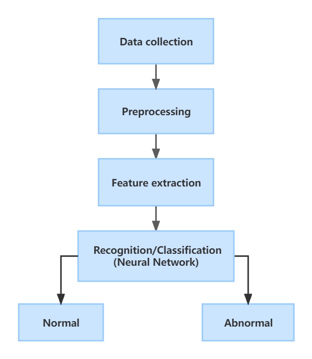
The models for brain tumour detection typically involve four phases (Figure 1). The first phase in the process is to collect accurate image data by sensors. The second phase is picture preprocessing, which includes improving the quality of the picture, enhancing details, and reducing noise. The third phase is featuring extraction, using the matrix method to extract the required features in the picture as input parameters. Finally, the neural network is responsible for the classification and the final result.

Figure 1. The common procedure of the method used in the brain tumor recognition.
2.2. Data collection
The gathering of data was an essential component of the investigation being conducted into the application of artificial intelligence in neurosurgery. The information that was gathered included imaging data from MRI or CT images, but it also included information about the patient, their medical background, and the results of any surgical procedures.
The reliability and precision of the data were necessary for the accomplishment of the research objectives [6]. Researchers had the option of utilizing the Digital Imaging and Communications in Medicine (DICOM) standard for medical pictures as a means of ensuring the precision of the data that had been collected.
2.3. Data preprocessing
The preprocessing procedures include the use of filters that can both smooth and sharpen the image. On the other hand, the sharpening filter is used to improve images by sharpening and accentuating minute detail or by increasing fuzzy detail. The smoothing filter is used to eliminate or minimize noise. The findings from the experiments show that the segmented pictures have a high level of precision while taking a much less amount of time for calculation. In the field of medical image processing, brain tumor segmentation is a necessary step. It is absolutely necessary for the treatment of people struggling with these illnesses to obtain an early diagnosis of brain tumors. If the brain tumor is discovered at an earlier stage, the patient has a better chance of surviving the illness [7]. Manual segmentation is commonly used by experts since the diagnosis of brain tumors requires automated segmentation. This approach is labor-intensive, time-consuming, and presents several challenges.
2.4. Feature extraction
After the picture has been segmented and preprocessed to remove abnormalities and noise, the next step is to locate regions of interest that are likely to contain cancerous growths. After the image has been segmented, a huge number of statistical and textural components are extracted from it by Gray-Level Co-Occurrence Matrix methods (GLCM) and Space Grey Level Dependency (SGLD) matrix. These elements, Energy, Correlation, Contrast, and Entropyare then put to use to evaluate if the brain tumor depicted in the image is benign or malignant.
2.5. Neural network for recognition or classification
A Natural-inspired Deep Convolutional Neural Network called ResNet-152 Transfer Learning model is used to identify and categorize the brain images using CNN and transfer learning techniques [8]. After input-detecting training, feature maps which are created by classifier CNN convolution layer are delivered to the next convolution layer with down sampling process. The flat input vector network is then used to produce an added number using each output neuron. This count indicates whether the input vector is divided into normal and abnormal brain tumors.
The analysis of MRI images can also be done by using back propagation network. The back propagation network with sigmoid transfer function training, includes three layers input, hidden and output layer [9]. The weights and biases of ANN show the relative strengths of each data transfer connection between layers. Then the characteristics curve which is plotted by the calculation for Haralick’s features can detect the tumor.
2.6. Evaluation metrics
The use of neurosurgical operations might benefit from the increased precision, efficiency, and safety afforded by artificial intelligence. Both the research plan and the data that was examined played a role in deciding which statistical evaluations were performed. Chi-square tests, regression analysis, T-tests, and one-way analysis of variance were some of the more common types of statistical investigations (ANOVA).
Historical studies investigate the application of AI in neurosurgery by looking at information that was gathered in the past, such as patient records. Planned cohort studies capture data on a collection of individuals over time to evaluate the efficacy of artificial intelligence (AI) [10]. Patients in randomized controlled studies are arbitrarily allocated to either a group that uses artificial intelligence (AI) or a group that does not use AI in order to evaluate their outcomes.
3. Application and discussion
Recent efforts to employ AI in the process of diagnosing brain lesions have generated positive results. MRI scans and other medical pictures are able to be evaluated by AI-based systems, which can help physicians diagnose brain cancer and identify other types of brain abnormalities.
3.1. Advantages of AI in brain tumor detection
The application of artificial intelligence (AI) technology in the detection of brain tumors presents numerous advantages, including the ability to quickly and accurately evaluate vast amounts of data, enabling earlier detection and improved diagnosis [11, 12]. AI systems utilize machine learning algorithms to differentiate between various forms of brain lesions and healthy tissue. Furthermore, AI can aid in tracking the development of brain lesions over time, potentially predicting tumor growth and patient response to treatment. In addition, AI may enhance the precision, efficiency, and safety of neurosurgical procedures by facilitating 3D reconstructions of the patient's brain to identify potential risks. While AI has the potential to improve diagnosis and treatment planning, it should not replace the expertise of trained medical professionals and should only be used as a tool to aid in the diagnosis process.
3.2. Challenges of AI in brain tumor detection
Medical professionals currently face challenges in using artificial diagnostic tools for identifying brain cancer, particularly in early detection when tumors are still small and have not yet metastasized [13]. The lack of accuracy in these instruments remains a significant obstacle, and the development of an explainable AI system for brain tumor detection is also a major challenge. High-performance deep learning models with hundreds of millions of parameters lack transparency and accountability, making it difficult to protect patient autonomy and their right to know. Additionally, despite data processing and analysis, surgical operations remain a crucial aspect of treatment. Surgery for brainstem tumors carries a high risk of intraoperative bleeding and damage to brain nerves, even with minimized surgical risk. Manual surgery, in particular, is prone to risk.
3.3. Future improvement of AI in brain tumor detection
In the future, AI has the potential to improve tumor detection in the future by enhancing the sensitivity, specificity, and throughput of detection. One potential approach is the training of AI algorithms to accurately identify and classify various tumor types using various imaging modalities such as MRI, CT, and ultrasound. Additionally, research should focus on developing AI systems for early cancer detection to improve cancer patients' survival rates [14]. To foster interdisciplinary collaboration and increase the transparency of AI systems, developers, medical professionals, and legislators should pay more attention to the limitations of opaque algorithms in medical AI. Robotics and automation are also significant for future development, particularly the use of surgical robots driven by AI that can assist neurosurgeons in executing difficult procedures with greater accuracy, stability, and dexterity than human hands alone, reducing the possibility of damaging tissue or making mistakes.
4. Conclusion
This study examines the application of artificial intelligence (AI) technology in the detection of brain tumors. The research process involves several stages, including data collection, information analysis, convolutional neural network (CNN) model construction, and evaluation. The article further evaluates the benefits of using AI in the field of brain tumor detection and medical treatment. The current challenges faced by this field relate to accuracy, explainability, and surgical risk, which must be addressed in future developments. The focus should be on creating an AI brain tumor detection system that is both explainable and powerful, along with the development of a sensor that can provide more precise image data and AI surgical robots. Additionally, AI may impact the development of national standards and enhance the efficiency of brain tumor research and therapy, ultimately improving clinical outcomes for patients in the future.
References
[1]. Ayadi W et al 2021 Deep CNN for brain tumor classification Neural Processing Letters 53 pp 671-700
[2]. Amin J et al 2021 Brain tumor detection and classification using machine learning: a comprehensive survey Complex & Intelligent Systems pp 1-23
[3]. Jian A Sidong L and Antonio D I 2022 Artificial intelligence for survival prediction in brain tumors on neuroimaging Neurosurgery 91.1 pp 8-26
[4]. Philip A K et al 2023 Artificial Intelligence and Precision Medicine: A New Frontier for the Treatment of Brain Tumors Life 13.1 24
[5]. Deepak S and Ameer P M 2019 Brain tumor classification using deep CNN features via transfer learning Computers in biology and medicine 111 103345.
[6]. Segato A et al 2020 Artificial intelligence for brain diseases: A systematic review APL bioengineering 4(4) 041503
[7]. King M R 2023 The future of AI in medicine: A perspective from a chatbot Annals of Biomedical Engineering 51(2) pp 291-295
[8]. A K K S et al 2022 A hybrid deep CNN-Cov-19-Res-Net Transfer learning architype for an enhanced Brain tumor Detection and Classification scheme in medical image processing Biomedical Signal Processing and Control 76
[9]. Abdalla H E M et al 2018 Brain Tumor Detection by using Artificial Neural Network," 2018 International Conference on Computer, Control, Electrical, and Electronics Engineering (ICCCEEE), Khartoum, Sudan pp 1-6
[10]. Palmisciano P 2020 Attitudes of patients and their relatives toward artificial intelligence in neurosurgery World neurosurgery 138 pp e627-e633
[11]. Abd-Ellah M K et al 2019 A review on brain tumor diagnosis from MRI images: Practical implications, key achievements, and lessons learned. Magnetic resonance imaging 61 pp 300-318
[12]. Yu Q et al 2019 Semantic segmentation of intracranial hemorrhages in head CT scans 2019 IEEE 10th International Conference on Software Engineering and Service Science (ICSESS). IEEE pp 112-115
[13]. Srinivas C et al 2022 Deep transfer learning approaches in performance analysis of brain tumor classification using MRI images Journal of Healthcare Engineering
[14]. Woźniak M 2021 Deep neural network correlation learning mechanism for CT brain tumor detection Neural Computing and Applications pp 1-16
Cite this article
Han,Z. (2023). The application and challenges of artificial intelligence in brain tumor recognition. Applied and Computational Engineering,17,17-22.
Data availability
The datasets used and/or analyzed during the current study will be available from the authors upon reasonable request.
Disclaimer/Publisher's Note
The statements, opinions and data contained in all publications are solely those of the individual author(s) and contributor(s) and not of EWA Publishing and/or the editor(s). EWA Publishing and/or the editor(s) disclaim responsibility for any injury to people or property resulting from any ideas, methods, instructions or products referred to in the content.
About volume
Volume title: Proceedings of the 5th International Conference on Computing and Data Science
© 2024 by the author(s). Licensee EWA Publishing, Oxford, UK. This article is an open access article distributed under the terms and
conditions of the Creative Commons Attribution (CC BY) license. Authors who
publish this series agree to the following terms:
1. Authors retain copyright and grant the series right of first publication with the work simultaneously licensed under a Creative Commons
Attribution License that allows others to share the work with an acknowledgment of the work's authorship and initial publication in this
series.
2. Authors are able to enter into separate, additional contractual arrangements for the non-exclusive distribution of the series's published
version of the work (e.g., post it to an institutional repository or publish it in a book), with an acknowledgment of its initial
publication in this series.
3. Authors are permitted and encouraged to post their work online (e.g., in institutional repositories or on their website) prior to and
during the submission process, as it can lead to productive exchanges, as well as earlier and greater citation of published work (See
Open access policy for details).
References
[1]. Ayadi W et al 2021 Deep CNN for brain tumor classification Neural Processing Letters 53 pp 671-700
[2]. Amin J et al 2021 Brain tumor detection and classification using machine learning: a comprehensive survey Complex & Intelligent Systems pp 1-23
[3]. Jian A Sidong L and Antonio D I 2022 Artificial intelligence for survival prediction in brain tumors on neuroimaging Neurosurgery 91.1 pp 8-26
[4]. Philip A K et al 2023 Artificial Intelligence and Precision Medicine: A New Frontier for the Treatment of Brain Tumors Life 13.1 24
[5]. Deepak S and Ameer P M 2019 Brain tumor classification using deep CNN features via transfer learning Computers in biology and medicine 111 103345.
[6]. Segato A et al 2020 Artificial intelligence for brain diseases: A systematic review APL bioengineering 4(4) 041503
[7]. King M R 2023 The future of AI in medicine: A perspective from a chatbot Annals of Biomedical Engineering 51(2) pp 291-295
[8]. A K K S et al 2022 A hybrid deep CNN-Cov-19-Res-Net Transfer learning architype for an enhanced Brain tumor Detection and Classification scheme in medical image processing Biomedical Signal Processing and Control 76
[9]. Abdalla H E M et al 2018 Brain Tumor Detection by using Artificial Neural Network," 2018 International Conference on Computer, Control, Electrical, and Electronics Engineering (ICCCEEE), Khartoum, Sudan pp 1-6
[10]. Palmisciano P 2020 Attitudes of patients and their relatives toward artificial intelligence in neurosurgery World neurosurgery 138 pp e627-e633
[11]. Abd-Ellah M K et al 2019 A review on brain tumor diagnosis from MRI images: Practical implications, key achievements, and lessons learned. Magnetic resonance imaging 61 pp 300-318
[12]. Yu Q et al 2019 Semantic segmentation of intracranial hemorrhages in head CT scans 2019 IEEE 10th International Conference on Software Engineering and Service Science (ICSESS). IEEE pp 112-115
[13]. Srinivas C et al 2022 Deep transfer learning approaches in performance analysis of brain tumor classification using MRI images Journal of Healthcare Engineering
[14]. Woźniak M 2021 Deep neural network correlation learning mechanism for CT brain tumor detection Neural Computing and Applications pp 1-16


