


1. Introduction
Extracellular vesicles are nanometer-sized, highly heterogeneous spherical solid bilayer proteolipid vesicles that are released into the extracellular space by virtually all different kinds of cells, including all different kinds of cells [1]. Extracellular bodies are a kind of substances with physiological functions, such as proteins, lipids, nucleic acids and so on. they are excreted in large quantities through their own physiological activities and enter into different organisms [2]. Extracellular vesicles consist of heterogeneous vesicles with different biological origins, compositions, and biological properties, including exosomes, microvesicles, and apoptotic cells [3,4]. Cells rely on extracellular vesicles to interact with the extracellular environment, and secretion of extracellular vesicles plays an important role in intercellular communication [5]. Extracellular bodies play an important role in regulating immune response (such as inherent and adaptive), angiogenesis, blood coagulation and miRNAs transmission [2,4]. In addition, EV is also involved in the pathogenesis of many diseases, such as tumorigenesis, growth and progression, and tumor invasion and metastasis is the key link of tumor treatment [1].
The contents of extracellular vesicles are expected to discover liquid biomarkers for prostate, kidney, and bladder cancers [6]. In addition, EV is also a medium that can help maintain the stability of the intra-articular environment [7]. Among them, EV microRNAs may be important in maintaining cardiovascular homeostasis and can coordinate the maintenance of cardiovascular homeostasis [8]. Cardiovascular diseases, including coronary heart disease, stroke, hypertension and peripheral artery disease, are not only the major economic burden on human health and society, but also the largest cause of cardiovascular disease morbidity and mortality worldwide [9]. EVS has a certain guiding significance for the occurrence and development of heart disease, and for clinical diagnosis, treatment and monitoring of heart disease [10](see Figure 1).
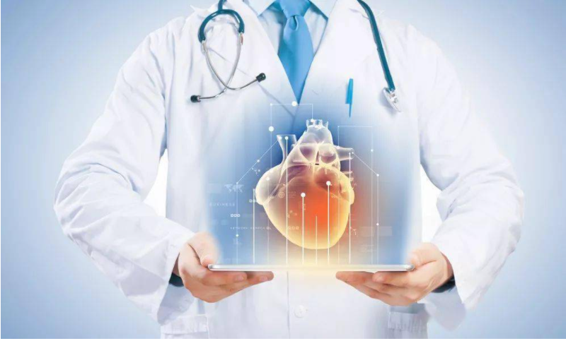
Figure 1. The Cardiovascular Health and Disease.
2. EV mirna as a CVD biomarker
MiRNAs, which is specifically expressed in intestinal epithelial cells, can be used as a prognostic and diagnostic marker in many diseases [11]. In 2007, Jan L€otvall and other studies showed that mutant RNA such as miRNAs and mRNAs can be mediated by the host in different hosts. In 2010, it was reported that transitive miRNAs could function in receiving cells [1]. Ev-mirna may be a predictor or indicator of early CVD detection. Deddens et al. noted that EV released into the circulation from a damaged heart contained mirna and proposed that EV mirna could be used as an early biomarker of cardiac injury. EV miRNAs can be used as an early diagnostic index of acute heart failure. Matsumoto et al. identified three p53-responsive mirna, which predicted the development of HF one year after acute myocardial infarction, by screening extensively for mirna in serum EVs. In addition, non-coding rna (ncRNA), especially circular rna (circRNA), which is carried in large quantities by EVs, has been recognized as a potential biomarker [11].
EV mirna has also been associated with cardiovascular risk factors (i.e., exposure to particulate matter, diabetes, dyslipidemia, obesity, MetS). Alterations in EV miRNA from particulate matter exposure to air pollution have been associated with elevated blood pressure and coagulation status, and have been shown to increase the risk of cardiovascular disease, as well as cardiovascular morbidity and mortality. It has been demonstrated that the transfer of miR-320 from cardiomyocytes to endothelial cells via EV has an inhibitory effect on myocardial angiogenesis. Down-regulated levels of both miRNAs in plasma EVs of patients with familial hypercholesterolemia (FH) and elevated levels of miR-130a resulted in decreased coronary atherosclerosis in patients with CAD, suggesting its use as a potential biomarker for CAD. Excessive obesity is another crucial risk factor for CVD, and EV mirna has been proposed as an early biomarker for predicting CVD events in obese patients. After coronary artery bypass grafting (CABG), EVs expresses a large amount of EVs miRNAs in circulation, which is closely related to coronary artery intima injury and can be used as a marker of ischemia-reperfusion injury in patients with coronary artery disease [12].
2.1. EV proteins as CVD prognostic biomarkers
The expression of virus (EV) is affected by various physiological and pathological factors. The protein profile may have changed in the disease’s early stages, making the content a potential early biomarker. EV proteins are considered as prognostic biomarkers for cardiovascular events. It has been shown that increased circulating CD31/membrane link protein 5-positive EV has an independent predictive effect on cardiovascular risk in patients with stable coronary atherosclerosis and that its release of high levels of EVs correlates with higher mortality rates and higher need for hemodialysis due to CVD [13].
2.2. EVs lipids as CVD prognostic biomarkers
The amount of lipids in EVs may be associated with atherosclerosis once these lipid accumulations are associated with toll-like receptor-mediated macrophage foam cell formation and apoptosis, leading to atherosclerosis. EV is rich in arachidonic acid, which can be secreted by activated platelets and stimulate the production of thrombin, thus accelerating the formation of thrombus [13].
2.3. EV counting as a biomarker of CVD
EV quantification has potential applications as a biomarker for diagnosis and therapeutic monitoring. About 70% of EV in normal people’s blood is secreted by platelets. Therefore, the EV in circulation is dominated by activated platelets, namely p-EV [11]. It has been shown that platelet-derived EV (p-EV) counts are associated with CVD. Under conditions of platelet-derived EV (p-EV) activation, such as myocardial infarction, there is an increased release of circulating EV in the plasma. In patients with ACS, the EV value increases, resulting in an ischemic pressure response. The number of EV also increased significantly in STEMI patients. In addition, EV increases rapidly in circulation after pathological stimulation. Deddens et al. found that the results show that this method can detect plasma vehicles quickly. Previous work found that EV increased within 1 hour after MI. The number of EVs may be an essential indicator for differentiating the severity of heart failure. The increase in the number of EVs derived from circulating endothelial cells is related to cardiac insufficiency, cardiac insufficiency, and so on. The concentration of EVS in patients with cardiac insufficiency was also higher than that in healthy people [13].
3. Conclusion
The cardioprotective effect of EV, especially the expression of miRNAs in cardiomyocytes, makes it a potential target for cardiomyocytes. EV proteins and lipids have been recognized as prognostic biomarkers of cardiovascular events. Quantification of EVs has potential applications as a biomarker for diagnosis and therapeutic monitoring. Exogenous or exogenous EV in blood and blood of EV has been proved to improve cardiac function and improve cardiac function, but its role in clinical practice remains to be further explored [11].
References
[1]. Chen, Z., Larregina, A. T., & Morelli, A. E. (2019). Impact of extracellular vesicles on innate immunity. Curr Opin Organ Transplant, 24(6), 670-678. doi:10.1097/MOT.0000000000000701
[2]. Chong, S. Y., Lee, C. K., Huang, C., Ou, Y. H., Charles, C. J., Richards, A. M., Wang, J. W. (2019). Extracellular Vesicles in Cardiovascular Diseases: Alternative Biomarker Sources, Therapeutic Agents, and Drug Delivery Carriers. Int J Mol Sci, 20(13). doi:10.3390/ijms20133272
[3]. de Freitas, R. C. C., Hirata, R. D. C., Hirata, M. H., & Aikawa, E. (2021). Circulating Extracellular Vesicles As Biomarkers and Drug Delivery Vehicles in Cardiovascular Diseases. Biomolecules, 11(3). doi:10.3390/biom11030388
[4]. Femmino, S., Penna, C., Margarita, S., Comita, S., Brizzi, M. F., & Pagliaro, P. (2020). Extracellular vesicles and cardiovascular system: Biomarkers and Cardioprotective Effectors. Vascul Pharmacol, 135, 106790. doi:10.1016/j.vph.2020.106790
[5]. Hutcheson, J. D., & Aikawa, E. (2018). Extracellular vesicles in cardiovascular homeostasis and disease. Curr Opin Cardiol, 33(3), 290-297. doi:10.1097/HCO.0000000000000510
[6]. Jansen, F., Nickenig, G., & Werner, N. (2017). Extracellular Vesicles in Cardiovascular Disease: Potential Applications in Diagnosis, Prognosis, and Epidemiology. Circ Res, 120(10), 1649-1657. doi:10.1161/CIRCRESAHA.117.310752
[7]. Kim, O. Y., Lee, J., & Gho, Y. S. (2017). Extracellular vesicle mimetics: Novel alternatives to extracellular vesicle-based theranostics, drug delivery, and vaccines. Semin Cell Dev Biol, 67, 74-82. doi:10.1016/j.semcdb.2016.12.001
[8]. Marin, D., & Scott, R. T., Jr. (2018). Extracellular vesicles: a promising tool for assessment of embryonic competence. Curr Opin Obstet Gynecol, 30(3), 171-178. doi:10.1097/GCO.0000000000000458
[9]. Miyaki, S., & Lotz, M. K. (2018). Extracellular vesicles in cartilage homeostasis and osteoarthritis. Curr Opin Rheumatol, 30(1), 129-135. doi:10.1097/BOR.0000000000000454
[10]. Osteikoetxea, X., Nemeth, A., Sodar, B. W., Vukman, K. V., & Buzas, E. I. (2016). Extracellular vesicles in cardiovascular disease: are they Jedi or Sith? J Physiol, 594(11), 2881-2894. doi:10.1113/JP271336
[11]. Pfeifer, P., Werner, N., & Jansen, F. (2015). Role and Function of MicroRNAs in Extracellular Vesicles in Cardiovascular Biology. Biomed Res Int, 2015, 161393. doi:10.1155/2015/161393
[12]. Urabe, F., Kosaka, N., Kimura, T., Egawa, S., & Ochiya, T. (2018). Extracellular vesicles: Toward a clinical application in urological cancer treatment. Int J Urol, 25(6), 533-543. doi:10.1111/iju.13594
[13]. Yang, X., Song, X., Li, Z., Liu, N., Yan, Y., & Liu, B. (2021). Crosstalk between extracellular vesicles and autophagy in cardiovascular pathophysiology. Pharmacol Res, 172, 105628. doi:10.1016/j.phrs.2021.105628
Cite this article
Cao,S. (2024). EVs as biomarkers of cardiovascular disease. Theoretical and Natural Science,46,61-64.
Data availability
The datasets used and/or analyzed during the current study will be available from the authors upon reasonable request.
Disclaimer/Publisher's Note
The statements, opinions and data contained in all publications are solely those of the individual author(s) and contributor(s) and not of EWA Publishing and/or the editor(s). EWA Publishing and/or the editor(s) disclaim responsibility for any injury to people or property resulting from any ideas, methods, instructions or products referred to in the content.
About volume
Volume title: Proceedings of the 2nd International Conference on Modern Medicine and Global Health
© 2024 by the author(s). Licensee EWA Publishing, Oxford, UK. This article is an open access article distributed under the terms and
conditions of the Creative Commons Attribution (CC BY) license. Authors who
publish this series agree to the following terms:
1. Authors retain copyright and grant the series right of first publication with the work simultaneously licensed under a Creative Commons
Attribution License that allows others to share the work with an acknowledgment of the work's authorship and initial publication in this
series.
2. Authors are able to enter into separate, additional contractual arrangements for the non-exclusive distribution of the series's published
version of the work (e.g., post it to an institutional repository or publish it in a book), with an acknowledgment of its initial
publication in this series.
3. Authors are permitted and encouraged to post their work online (e.g., in institutional repositories or on their website) prior to and
during the submission process, as it can lead to productive exchanges, as well as earlier and greater citation of published work (See
Open access policy for details).
References
[1]. Chen, Z., Larregina, A. T., & Morelli, A. E. (2019). Impact of extracellular vesicles on innate immunity. Curr Opin Organ Transplant, 24(6), 670-678. doi:10.1097/MOT.0000000000000701
[2]. Chong, S. Y., Lee, C. K., Huang, C., Ou, Y. H., Charles, C. J., Richards, A. M., Wang, J. W. (2019). Extracellular Vesicles in Cardiovascular Diseases: Alternative Biomarker Sources, Therapeutic Agents, and Drug Delivery Carriers. Int J Mol Sci, 20(13). doi:10.3390/ijms20133272
[3]. de Freitas, R. C. C., Hirata, R. D. C., Hirata, M. H., & Aikawa, E. (2021). Circulating Extracellular Vesicles As Biomarkers and Drug Delivery Vehicles in Cardiovascular Diseases. Biomolecules, 11(3). doi:10.3390/biom11030388
[4]. Femmino, S., Penna, C., Margarita, S., Comita, S., Brizzi, M. F., & Pagliaro, P. (2020). Extracellular vesicles and cardiovascular system: Biomarkers and Cardioprotective Effectors. Vascul Pharmacol, 135, 106790. doi:10.1016/j.vph.2020.106790
[5]. Hutcheson, J. D., & Aikawa, E. (2018). Extracellular vesicles in cardiovascular homeostasis and disease. Curr Opin Cardiol, 33(3), 290-297. doi:10.1097/HCO.0000000000000510
[6]. Jansen, F., Nickenig, G., & Werner, N. (2017). Extracellular Vesicles in Cardiovascular Disease: Potential Applications in Diagnosis, Prognosis, and Epidemiology. Circ Res, 120(10), 1649-1657. doi:10.1161/CIRCRESAHA.117.310752
[7]. Kim, O. Y., Lee, J., & Gho, Y. S. (2017). Extracellular vesicle mimetics: Novel alternatives to extracellular vesicle-based theranostics, drug delivery, and vaccines. Semin Cell Dev Biol, 67, 74-82. doi:10.1016/j.semcdb.2016.12.001
[8]. Marin, D., & Scott, R. T., Jr. (2018). Extracellular vesicles: a promising tool for assessment of embryonic competence. Curr Opin Obstet Gynecol, 30(3), 171-178. doi:10.1097/GCO.0000000000000458
[9]. Miyaki, S., & Lotz, M. K. (2018). Extracellular vesicles in cartilage homeostasis and osteoarthritis. Curr Opin Rheumatol, 30(1), 129-135. doi:10.1097/BOR.0000000000000454
[10]. Osteikoetxea, X., Nemeth, A., Sodar, B. W., Vukman, K. V., & Buzas, E. I. (2016). Extracellular vesicles in cardiovascular disease: are they Jedi or Sith? J Physiol, 594(11), 2881-2894. doi:10.1113/JP271336
[11]. Pfeifer, P., Werner, N., & Jansen, F. (2015). Role and Function of MicroRNAs in Extracellular Vesicles in Cardiovascular Biology. Biomed Res Int, 2015, 161393. doi:10.1155/2015/161393
[12]. Urabe, F., Kosaka, N., Kimura, T., Egawa, S., & Ochiya, T. (2018). Extracellular vesicles: Toward a clinical application in urological cancer treatment. Int J Urol, 25(6), 533-543. doi:10.1111/iju.13594
[13]. Yang, X., Song, X., Li, Z., Liu, N., Yan, Y., & Liu, B. (2021). Crosstalk between extracellular vesicles and autophagy in cardiovascular pathophysiology. Pharmacol Res, 172, 105628. doi:10.1016/j.phrs.2021.105628