


1. Introduction
Gliomas are a group of malignant intracranial tumors that occur mostly in the central nervous system and account for approximately 50% of all brain tumor patients [1-2]. According to clinical grading, gliomas are classified into grades I to IV, and their clinical manifestations are closely related to the site of occurrence and the size of the tumor. Grades I and II are low-grade gliomas, while grades III and IV are high-grade gliomas. Treatment options for gliomas vary with different grades, and accurate grading is clinically important for planning treatment and improving prognosis. Gliomas are typically heterogeneous, and thus histologic samples obtained at biopsy may be subject to sampling error, and intracranial biopsy is relatively difficult [3]. Therefore, it is often graded using imaging techniques. This paper discusses the image characteristics of Magnetic Resonance Imaging (MRI) and Computed Tomography (CT) techniques to determine their value in the staging of gliomas. This paper reviews previous studies in China and abroad that applied imaging methods to apply grading to gliomas and summarizes the main techniques of MRI and CT and their clinical effects by using the literature analysis method. This study organizes the current stage of medical imaging technology in the field of glioma grading as well as the latest progress and trends, which can give some references for Related researchers.
2. Imaging Grading of Gliomas
2.1. Gliomas on MRI
MRI is an important imaging technique for the clinical diagnosis of gliomas, and several sequences of MRI such as Diffusion Weighted Imaging (DWI) and Perfusion Weighted Imaging (PWI) can be applied in the grading diagnosis of gliomas. DWI is able to detect the diffusive movement of water molecules in tissues, which can be responded to by measuring the Approximate Diffusion Coefficient (ADC). Some studies have shown that, due to the high cytoplasmic nature of the tumor, high-grade gliomas tend to exhibit lower ADC (see Figure 1) [4]. PWI is the use of rapid enhancement scanning to determine perfusion by contrast signal intensity. High-grade gliomas will show aggressive growth, so the higher the grade of the glioma, the greater the proliferation of blood vessels [5]. Wang et al used the parameter Relative Cerebral Blood Volume (RCBV) in PWI as an observational indicator, it can be seen that in the parenchymal region of the tumor, the RCBV of low-grade gliomas was significantly lower than that of high-grade gliomas [6]; in the edematous region surrounding the tumor, the RCBV of low-grade gliomas was significantly lower than that of high-grade gliomas.
This shows that MRI is effective in preoperative grading of gliomas.
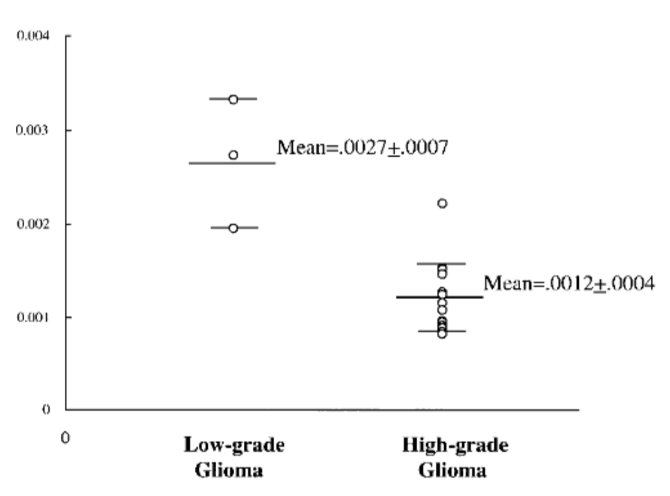
Figure 1. Apparent diffusion coefficient (ADC) value versusgrading of gliomas.
The ADC value is higher in the high-gradegliomas than in the low-grade gliomas. The difference betweenthe means is statistically significant (P,0.001) [4].
2.2. Gliomas on CT
CT is also a common imaging technique used clinically to diagnose gliomas because CT is better at showing hemorrhages and calcifications within the lesion. Among them, multilayer spiral CT perfusion imaging has high temporal resolution, spatial resolution and density resolution, which can effectively detect the functional status of brain tissue, so it can be used for diagnosis and grading of gliomas in the brain [7]. The main parameters of CT perfusion imaging include Blood Flow (BF), Blood Volume (BV), and surface permeability coefficient (PS). According to Zhang et al, and Liu et al, the higher the pathological grade, the higher the BF, BV, and PS values in the parenchymal region of gliomas, and different grades of gliomas can be distinguished based on these parameters [8-9].
3. Advantages And Disadvantages of MRI Vs. Ct
3.1. Advantages and disadvantages of MRI
MRI is radiation-free and does not cause radioactive damage to brain tissue, and it has high soft tissue resolution and obvious contrast of different tissues, which can intuitively determine the structure within the tumor, and it also has the advantages of being able to be multi-directional, multi-sequence imaging and three-dimensional imaging. However, MRI examination takes a longer time and has more contraindications (including claustrophobia, metal implants, etc.), especially unsuitable for patients with acute and critical diseases.
3.2. Advantages and disadvantages of CT
CT scanning is fast and has a wide range of indications. It has the advantage of responding to characteristic density changes such as calcifications, hemorrhages, and cystic lesions, but there are limitations in its application to diffusely growing tumors like gliomas, which may be subject to errors due to changes in normal anatomy caused by bands of edema and compression by the tumor [10]. In addition, CT examinations can be compromised by ionizing radiation.
4. Combination of MRI and CT
Currently, clinical diagnosis and grading of gliomas are mainly performed by MRI and CT, but the validity of the imaging methods is still controversial. In summary, it can be seen that MRI and CT are not mutually exclusive techniques per se, and they can be used synergistically to further diagnose gliomas. According to existing studies, the combined use of CT and MRI techniques can provide complementary information to more accurately determine the extent of the tumor, the demarcation from normal tissue, and the precise structures and alterations within the tumor. According to Wu, the diagnostic ability of patients using CT combined with MRI was better than that of CT alone or MRI alone, and the sensitivity and specificity of the combined application were relatively high, reaching 97% and 100%[11]; according to Long et al, the diagnostic sensitivity of CT alone for the diagnosis of gliomas (80.00%), and the specificity of MRI alone for the diagnosis of gliomas (82.22%) were significantly lower than that of CT combined with MRI (P < 0.05), and the diagnostic value for gliomas was significantly lower than that of CT combined with MRI (P < 0.05) [12].
5. Conclusion
This paper found that both MRI and CT have techniques that can be used individually to grade gliomas with good results, while the combined application of MRI and CT has high sensitivity and specificity for the diagnosis and grading of gliomas, and deserves to be widely used in clinical practice. There is some room for improvement in this paper: a. This paper does not discuss all imaging techniques that can be used to grade gliomas, and only roughly summarizes popular and commonly used techniques; b. Most of the literature included are retrospective studies, the current status of the subjects is unknown, and a certain number of prospective studies are needed to more accurately validate the reliability and usefulness of the imaging methods in clinical practice. Future studies could focus on the combination of multiple imaging techniques for glioma grading to improve the speed of examination and diagnostic quality.
References
[1]. Ma Qingyou, Geng Qilong & Wang Fafen. (2022). Application Value of Magnetic Resonance Imaging in Differential Diagnosis of Glioma. Chinese Community Doctors(16),96-98. doi:CNKI:SUN:XCYS.0.2022-16-032.
[2]. Marsh, J. C., Wendt, J. A., Herskovic, A. M., Diaz, A., Gielda, B. T., & Byrne, R. W. (2012). High-grade glioma relationship to the neural stem cell compartment: a retrospective review of 104 cases. International journal of radiation oncology, biology, physics, 82(2), e159–e165. https://doi.org/10.1016/j.ijrobp.2010.08.036
[3]. Ryu, Y. J., Choi, S. H., Park, S. J., Yun, T. J., Kim, J. H., & Sohn, C. H. (2014). Glioma: application of whole-tumor texture analysis of diffusion-weighted imaging for the evaluation of tumor heterogeneity. PloS one, 9(9), e108335. https://doi.org/10.1371/ journal.pone.0108335
[4]. Sugahara, T., Korogi, Y., Kochi, M., Ikushima, I., Shigematu, Y., Hirai, T., Okuda, T., Liang, L., Ge, Y., Komohara, Y., Ushio, Y., & Takahashi, M. (1999). Usefulness of diffusion-weighted MRI with echo-planar technique in the evaluation of cellularity in gliomas. Journal of magnetic resonance imaging : JMRI, 9(1), 53–60. https://doi.org/10.1002/(sici)1522-2586 (199901)9:1<53::aid-jmri7>3.0.co;2-2
[5]. Li Yongming. (2020). The differential diagnostic value of magnetic resonance imaging in deep lesions of primary central nervous system lymphoma and high-grade glioma. Henan Medical Research(08), 1477-1478. doi:CNKI:SUN:HNYX.0.2020-08-070.
[6]. Wang Wei, Li Hong, Ji Peng & Tan Wengang. (2020). Analysis The Diagnostic Value of MR Perfusion Weighted Imaging for the Diagnosis of Gliomas and Brain Single Metastasis. Chinese Journal of CT and MRI(08),19-21. doi:CNKI:SUN:CTMR.0.2020-08-007.
[7]. Ma Xiuhua, Lv Furong, Lv Fajin, Xiao Zhibo, Huang Youxin, Li Xinyou... & Pan Jing. (2009). Evaluating rat model of acute cerebral ischemia-reperfusion by computed tomography perfusion imaging.. Acta Academiae Medicinae Militaris Tertiae (06),506-509. doi:CNKI:SUN:DSDX.0.2009-06-015.
[8]. Zhang Changfei, Du Fuchuan & Zhang Changkai. (2021).Feasibility that Multi-slice Spiral CT Perfusion Imaging is Used to Evaluate the Pathological Grade of Glioma. Chinese Journal of CT and MRI(09),22-24. doi:CNKI:SUN:CTMR.0.2021-09-043.
[9]. Liu Gang, Cui Guosheng, Sun Baoshan & Pan Liang. (2016). Application of Multi-slice Spiral CT Perfusion imaging in Cerebral Glioma Pathological grade. Chinese And Foreign Medical Research(06),61-62. doi:10.14033/j.cnki.cfmr.2016.6.033.
[10]. Bai Zhenglu, Li Jun & Tian Shuchang. (2020). Application of CT-MR Image Fusion in Postoperative Radiotherapy for Gliomas. China Medical Devices (12),20-23. doi:CNKI:SUN:YLSX.0.2020-12-006.
[11]. Wu Baolai. (2021). Application of CT combined with magnetic resonance imaging in preoperative diagnosis of glioma. Chinese Remedies & Clinics (14), 2472-2474. doi:CNKI:SUN:YWLC.0.2021-14-022.
[12]. Long Chunqin, Zhou Zhiqiang, He Wenjun, Zhang Zhoubing & Zhang Dawei. (2020). Application of CT Combined with MRI in Preoperative Diagnosis of Glioma. Chinese Journal of CT and MRI(06),9-11+21. doi:CNKI:SUN:CTMR.0.2020-06-003.
Cite this article
Wang,Z. (2023). Analysis of the clinical display effect of imaging techniques on the staging of gliomas. Theoretical and Natural Science,17,9-12.
Data availability
The datasets used and/or analyzed during the current study will be available from the authors upon reasonable request.
Disclaimer/Publisher's Note
The statements, opinions and data contained in all publications are solely those of the individual author(s) and contributor(s) and not of EWA Publishing and/or the editor(s). EWA Publishing and/or the editor(s) disclaim responsibility for any injury to people or property resulting from any ideas, methods, instructions or products referred to in the content.
About volume
Volume title: Proceedings of the 2nd International Conference on Modern Medicine and Global Health
© 2024 by the author(s). Licensee EWA Publishing, Oxford, UK. This article is an open access article distributed under the terms and
conditions of the Creative Commons Attribution (CC BY) license. Authors who
publish this series agree to the following terms:
1. Authors retain copyright and grant the series right of first publication with the work simultaneously licensed under a Creative Commons
Attribution License that allows others to share the work with an acknowledgment of the work's authorship and initial publication in this
series.
2. Authors are able to enter into separate, additional contractual arrangements for the non-exclusive distribution of the series's published
version of the work (e.g., post it to an institutional repository or publish it in a book), with an acknowledgment of its initial
publication in this series.
3. Authors are permitted and encouraged to post their work online (e.g., in institutional repositories or on their website) prior to and
during the submission process, as it can lead to productive exchanges, as well as earlier and greater citation of published work (See
Open access policy for details).
References
[1]. Ma Qingyou, Geng Qilong & Wang Fafen. (2022). Application Value of Magnetic Resonance Imaging in Differential Diagnosis of Glioma. Chinese Community Doctors(16),96-98. doi:CNKI:SUN:XCYS.0.2022-16-032.
[2]. Marsh, J. C., Wendt, J. A., Herskovic, A. M., Diaz, A., Gielda, B. T., & Byrne, R. W. (2012). High-grade glioma relationship to the neural stem cell compartment: a retrospective review of 104 cases. International journal of radiation oncology, biology, physics, 82(2), e159–e165. https://doi.org/10.1016/j.ijrobp.2010.08.036
[3]. Ryu, Y. J., Choi, S. H., Park, S. J., Yun, T. J., Kim, J. H., & Sohn, C. H. (2014). Glioma: application of whole-tumor texture analysis of diffusion-weighted imaging for the evaluation of tumor heterogeneity. PloS one, 9(9), e108335. https://doi.org/10.1371/ journal.pone.0108335
[4]. Sugahara, T., Korogi, Y., Kochi, M., Ikushima, I., Shigematu, Y., Hirai, T., Okuda, T., Liang, L., Ge, Y., Komohara, Y., Ushio, Y., & Takahashi, M. (1999). Usefulness of diffusion-weighted MRI with echo-planar technique in the evaluation of cellularity in gliomas. Journal of magnetic resonance imaging : JMRI, 9(1), 53–60. https://doi.org/10.1002/(sici)1522-2586 (199901)9:1<53::aid-jmri7>3.0.co;2-2
[5]. Li Yongming. (2020). The differential diagnostic value of magnetic resonance imaging in deep lesions of primary central nervous system lymphoma and high-grade glioma. Henan Medical Research(08), 1477-1478. doi:CNKI:SUN:HNYX.0.2020-08-070.
[6]. Wang Wei, Li Hong, Ji Peng & Tan Wengang. (2020). Analysis The Diagnostic Value of MR Perfusion Weighted Imaging for the Diagnosis of Gliomas and Brain Single Metastasis. Chinese Journal of CT and MRI(08),19-21. doi:CNKI:SUN:CTMR.0.2020-08-007.
[7]. Ma Xiuhua, Lv Furong, Lv Fajin, Xiao Zhibo, Huang Youxin, Li Xinyou... & Pan Jing. (2009). Evaluating rat model of acute cerebral ischemia-reperfusion by computed tomography perfusion imaging.. Acta Academiae Medicinae Militaris Tertiae (06),506-509. doi:CNKI:SUN:DSDX.0.2009-06-015.
[8]. Zhang Changfei, Du Fuchuan & Zhang Changkai. (2021).Feasibility that Multi-slice Spiral CT Perfusion Imaging is Used to Evaluate the Pathological Grade of Glioma. Chinese Journal of CT and MRI(09),22-24. doi:CNKI:SUN:CTMR.0.2021-09-043.
[9]. Liu Gang, Cui Guosheng, Sun Baoshan & Pan Liang. (2016). Application of Multi-slice Spiral CT Perfusion imaging in Cerebral Glioma Pathological grade. Chinese And Foreign Medical Research(06),61-62. doi:10.14033/j.cnki.cfmr.2016.6.033.
[10]. Bai Zhenglu, Li Jun & Tian Shuchang. (2020). Application of CT-MR Image Fusion in Postoperative Radiotherapy for Gliomas. China Medical Devices (12),20-23. doi:CNKI:SUN:YLSX.0.2020-12-006.
[11]. Wu Baolai. (2021). Application of CT combined with magnetic resonance imaging in preoperative diagnosis of glioma. Chinese Remedies & Clinics (14), 2472-2474. doi:CNKI:SUN:YWLC.0.2021-14-022.
[12]. Long Chunqin, Zhou Zhiqiang, He Wenjun, Zhang Zhoubing & Zhang Dawei. (2020). Application of CT Combined with MRI in Preoperative Diagnosis of Glioma. Chinese Journal of CT and MRI(06),9-11+21. doi:CNKI:SUN:CTMR.0.2020-06-003.